. 2003 Jul;23(13):4586-97.
doi: 10.1128/MCB.23.13.4586-4597.2003.

The murine Nck SH2/SH3 adaptors are important for the development of mesoderm-derived embryonic structures and for regulating the cellular actin network

Affiliations

The murine Nck SH2/SH3 adaptors are important for the development of mesoderm-derived embryonic structures and for regulating the cellular actin network

Friedhelm Bladt et al. Mol Cell Biol. 2003 Jul.

Abstract

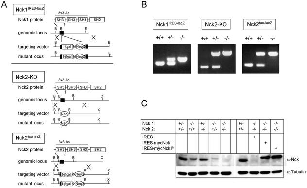
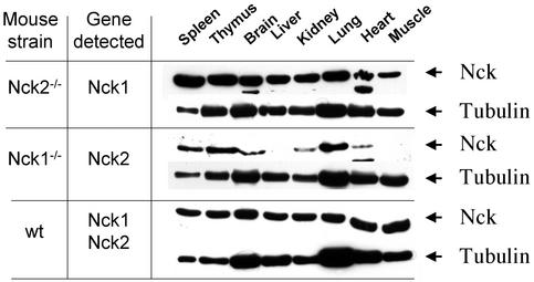
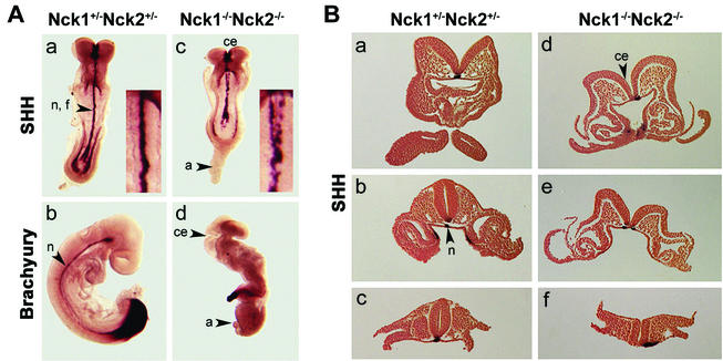
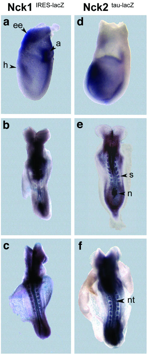
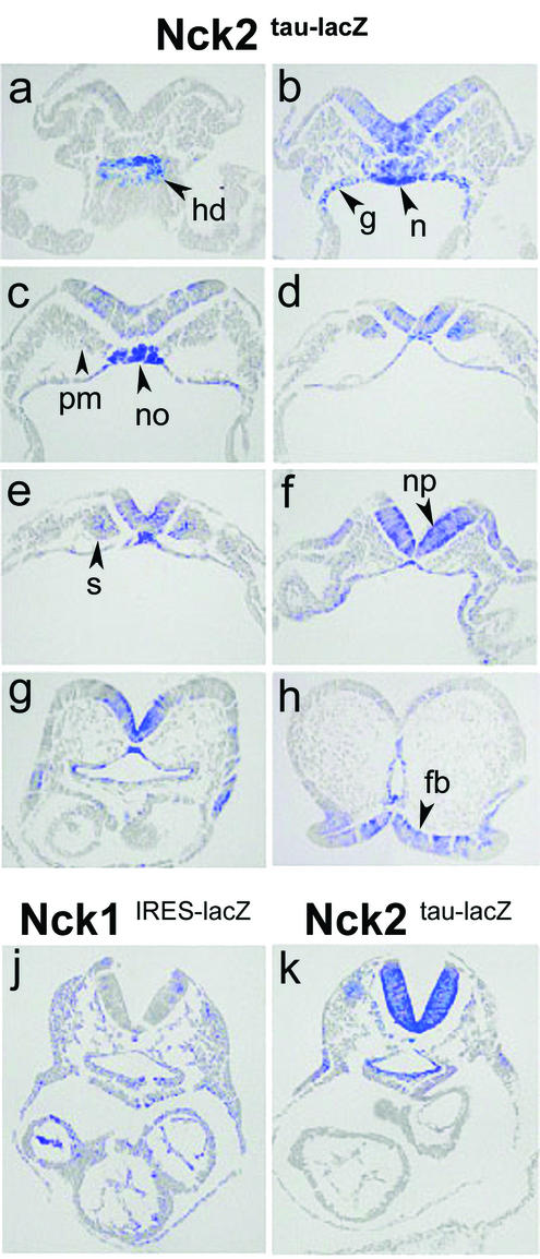
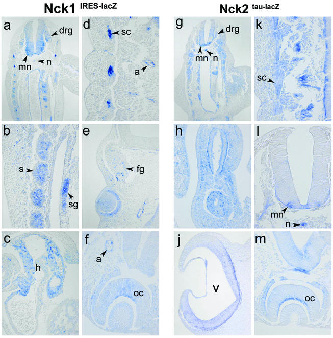
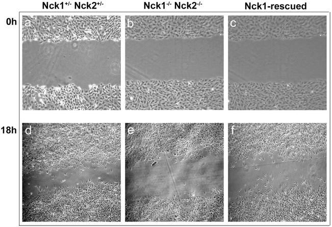
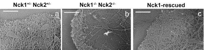
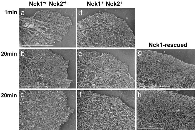
Mammalian Nck1 and Nck2 are closely related adaptor proteins that possess three SH3 domains, followed by an SH2 domain, and are implicated in coupling phosphotyrosine signals to polypeptides that regulate the actin cytoskeleton. However, the in vivo functions of Nck1 and Nck2 have not been defined. We have mutated the murine Nck1 and Nck2 genes and incorporated beta-galactosidase reporters into the mutant loci. In mouse embryos, the two Nck genes have broad and overlapping expression patterns. They are functionally redundant in the sense that mice deficient for either Nck1 or Nck2 are viable, whereas inactivation of both Nck1 and Nck2 results in profound defects in mesoderm-derived notochord and embryonic lethality at embryonic day 9.5. Fibroblast cell lines derived from Nck1(-/-) Nck2(-/-) embryos have defects in cell motility and in the organization of the lamellipodial actin network. These data suggest that the Nck SH2/SH3 adaptors have important functions in the development of mesodermal structures during embryogenesis, potentially linked to a role in cell movement and cytoskeletal organization.


Figures

FIG. 1.
FIG. 1.
Generation of Nck1- and Nck2-deficient mouse strains. (A) Schematic representation of the gene-targeting strategies used to mutate the Nck genes. The restriction maps of the Nck1 and Nck2 loci are indicated (B, BamHI; E, EcoRI; X, XbaI). The Nck1IRES-lacZ targeting vector included an IRES-lacZ reporter gene cassette. The Nck2tau-lacZ targeting vector comprised a tau-lacZ reporter gene fused in frame with the first coding exon. The protein region employed to raise the 3x3 polyclonal antibody used for Western blot analysis is indicated. (B) Genotypic analysis of the mutant mouse strains by PCR. Primer combinations are indicated in the Methods. The WT and mutant bands were, respectively, of 210 and 280 bp for Nck1, 250 and 610 bp for Nck2ko, and 170 and 280 bp for Nck2tau-lacZ mutant mouse strains. (C) Expression of the Nck genes was assessed by Western blotting of protein extracts of MEFs with the indicated genotypes. Western blots of protein extracts of mutant MEFs infected with IRES-EGFP and myc-tagged Nck1-IRES-EGFP retroviral vectors are also shown.
FIG. 2.
FIG. 2.
Differential pattern of expression of Nck1 and Nck2 in adult tissues. The expression of Nck1 and Nck2 was assessed in various tissue extracts from Nck1−/− Nck2−/− mutants and WT mice. Equivalent amounts of tissue extracts (determined by Bio-Rad assay) were probed with the 3x3 polyclonal antibody, recognizing the first SH3 domain of Nck1 and Nck2. Since this region was deleted in our targeting strategies, we used tissues from Nck2−/− and Nck1−/− mutants to assess the expression of Nck1 and Nck2, respectively. Membranes were stripped and reblotted with anti-β-tubulin antibody. Arrowheads mark the position of the indicated protein bands. The results are representative of two independent experiments.
FIG. 3.
FIG. 3.
Concomitant loss of Nck1 and Nck2 functions results in multiple morphological abnormalities and lack of axial rotation. (A) Shh and Brachyury staining of Nck1+/− Nck2+/− and Nck1−/− Nck2−/− embryos at E8.5 (a and c) and E9.5 (b and d). In Nck1+/− Nck2+/− embryos, Shh stained the notochord, floor plate, and endodermal cells of the hind- and foregut (a). In Nck1−/− Nck2−/− embryos, Shh staining could be detected in the notochord floorplate and endodermal cells. The notochord floorplate was bifurcated and disrupted along the posterior-anterior axis (c). Brachyury expression is evident in the tail bud and notochord of E9.5 control embryo (b). Nck1−/− Nck2−/− embryos (d) did not “turn,” and the allantois did not fuse with the chorion and acquired a balloon-like structure. The headfolds were not closed. Fragmentary brachyury staining was visible in the tail bud and along the posterior-anterior axis. (B) Sections from Nck1+/− Nck2+/− and Nck1−/− Nck2−/− mutant E8.5 embryos stained with Shh. In Nck1+/− Nck2+/− embryos, notochord (a to c), floor plate (a to c), and endodermal precursor cells (b) were positive for Shh. In mutant embryos, Shh staining revealed displacement beside the midline (d and f) and bifurcation (e) of notochord and floor plate. Abbreviations: n, notochord; f, floorplate; a, allantois; ce, cephalic folds.
FIG. 4.
FIG. 4.
Expression pattern of Nck1 and Nck2 at E7.5 and E8.5. β-Galactosidase activity was exhibited by E7.5 (a and d) and E8.5 (b, c, e, and f) embryos heterozygous for the Nck1IRES-lacZ and Nck2tau-lacZ loci. Although weak β-galactosidase activity was detected throughout both Nck1 and Nck2 heterozygous embryos, strong expression of both the Nck1 and Nck2 genes localized to the allantois (a), headfold (h), and extraembryonic (ee) tissues (panels a and d). At E8.5 both genes were expressed in the node (n), somites (s), and neural tube (nt; panels b, c, e, and f). In Nck1IRES-lacZ embryos, weak staining was also found in the yolk sac.
FIG. 5.
FIG. 5.
Comparative analysis of the expression pattern of Nck1 and Nck2 at E8.5. Transverse sections of X-Gal-stained heterozygous Nck2tau-lacZ and Nck1IRES-lacZ E8.5 embryos. In heterozygous Nck2tau-lacZembryos, β-galactosidase activity was detected in the caudal extremity of hindgut diverticulum (a); endodermal lining of hind-, mid-, and foregut (b to g); caudal region of notochord (b); notochordal plate (c); notochord (f and g); neural plate (b to f); neuroepithelium of prospective forebrain region (h); paraxial mesoderm (b and c); and somites (d and e). (j and k) Comparative analysis of similar transverse sections from homozygous Nck1IRES-lacZ and Nck2tau-lacZ mutants at E8.5 revealed expression of both genes in most tissues. However, whereasNck1 was expressed throughout the embryo, Nck2 exhibited a more restricted pattern of expression and was undetectable in the hearts of Nck2tau-lacZ embryos. Abbreviations: hd, hindgut diverticulum; g, hind-, mid-, and foregut; n, notochord; no, notochordal plate; np, neural plate; fb, forebrain; pm, paraxial mesoderm; s, somites.
FIG. 6.
FIG. 6.
Nck1 and Nck2 have highly overlapping expression patterns and are particularly prominent in the developing nervous system. Transverse sections of X-Gal-stained Nck1IRES-lacZ and Nck2tau-lacZ embryos at E10.5. Nck1 and Nck2 were highly expressed in neural structures such as motorneurons (a, g, k, and l), dorsal root ganglia (a and g), facial ganglia (e), sympathetic trunk ganglia (b), the inner layer of optic cup (f and m, future nervous layer of the retina), and the neuroepithelium of the ventricles (j). However, expression was also detected in nonneural structures such as the notochord (a, g, and l), somites (a, b, and g), sclerotome (a, d, and k), and arteries (d to f and m). Interestingly, the Nck proteins were differentially expressed in the arterial chamber of the heart (c), where the expression of Nck1, but not Nck2, could be detected. Abbreviations: m, motorneurons; drg, dorsal root ganglia; fg, facial ganglia; sg, sympathetic trunk ganglia; oc, optic cup; v, ventricle; n, notochord; s, somites; sc, sclerotome; a, arteries; h, heart.
FIG. 7.
FIG. 7.
Impaired motility of Nck1−/− Nck2−/− MEFs. Confluent monolayers of Nck1+/− Nck2+/−, Nck1−/− Nck2−/−, and Nck1-rescued MEFs were mechanically interrupted, and the inflicted wounds were photographed at the indicated time points (in hours). Nck1−/− Nck2−/− cells exhibited reduced ability to migrate into the wound compared to Nck1+/− Nck2+/− and Nck1-rescued MEFs.
FIG. 8.
FIG. 8.
Altered lamellipodium formation in Nck1−/− Nck2−/− MEFs. Platinum replica electron microscopy of lamellipodia of Nck1−/− Nck2+/−, Nck1−/− Nck2−/−, and Nck1-rescued MEFs, plated on fibronectin, was done. (a) In Nck1+/− Nck2+/− MEFs, a dense actin filament network of ca. 1 μm thick, the “leading edge,” protruded out of a finely branched actin filamentous meshwork. (b) In Nck1−/− Nck2−/− MEFs, the leading edge was shorter and irregularly shaped and exhibited, at the ultrastructural level, short actin filaments. Furthermore, in the area immediately behind the leading edge, the actin filaments were consistently much sparser and less branched. (c) In the Nck1-rescued MEFs, the leading edge was characterized by an actin filament network of density comparable to that of Nck1+/− Nck2+/− MEFs. Scale bar, 1 μm.
FIG. 9.
FIG. 9.
Actin network organization upon ATP recovery. Platinum replica electron microscopy of lamellipodia of Nck1+/− Nck2+/−, Nck1−/− Nck2−/−, and Nck1-rescued MEFs after a 1- or 20-min recovery period from ATP depletion was completed. One minute after sodium azide washout, the recovery of Nck1−/− Nck2−/− MEFs was comparable to that of Nck1+/− Nck2+/− and Nck1-rescued MEFs. In contrast, after 20 min, the recovery of Nck1−/− Nck2−/− MEFs was significantly impaired compared to that of Nck1+/− Nck2+/− and Nck1-rescued MEFs. Nck1−/− Nck2−/− MEFs had shorter filaments at the leading edge and less-branched filaments behind the leading edge.

References

    1. Anton, I. M., W. Lu, B. J. Mayer, N. Ramesh, and R. S. Geha. 1998. The Wiskott-Aldrich syndrome protein-interacting protein (WIP) binds to the adaptor protein Nck. J. Biol. Chem. 273:20992-20995. - PubMed
    1. Becker, E., U. Huynh-Do, S. Holland, T. Pawson, T. O. Daniel, and E. Y. Skolnik. 2000. Nck-interacting Ste20 kinase couples Eph receptors to c-Jun N-terminal kinase and integrin activation. Mol. Cell. Biol. 20:1537-1545. - PMC - PubMed
    1. Beddington, R. S. 1994. Induction of a second neural axis by the mouse node. Development 120:613-620. - PubMed
    1. Bokoch, G. M., Y. Wang, B. P. Bohl, M. A. Sells, L. A. Quilliam, and U. G. Knaus. 1996. Interaction of the Nck adapter protein with p21-activated kinase (PAK1). J. Biol. Chem. 271:25746-25749. - PubMed
    1. Brand-Saberi, B., and B. Christ. 2000. Evolution and development of distinct cell lineages derived from somites. Curr. Top. Dev. Biol. 48:1-42. - PubMed

Publication types

MeSH terms

LinkOut - more resources