. 2003 Jul;23(13):4637-48.
doi: 10.1128/MCB.23.13.4637-4648.2003.

Targeted disruption of Aldh1a1 (Raldh1) provides evidence for a complex mechanism of retinoic acid synthesis in the developing retina

Affiliations

Targeted disruption of Aldh1a1 (Raldh1) provides evidence for a complex mechanism of retinoic acid synthesis in the developing retina

Xiaohong Fan et al. Mol Cell Biol. 2003 Jul.

Abstract

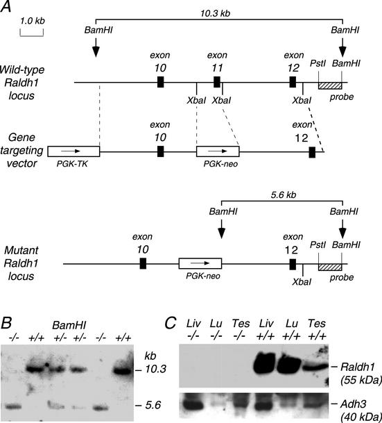
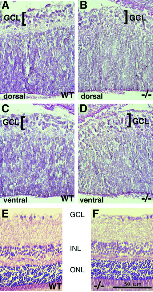
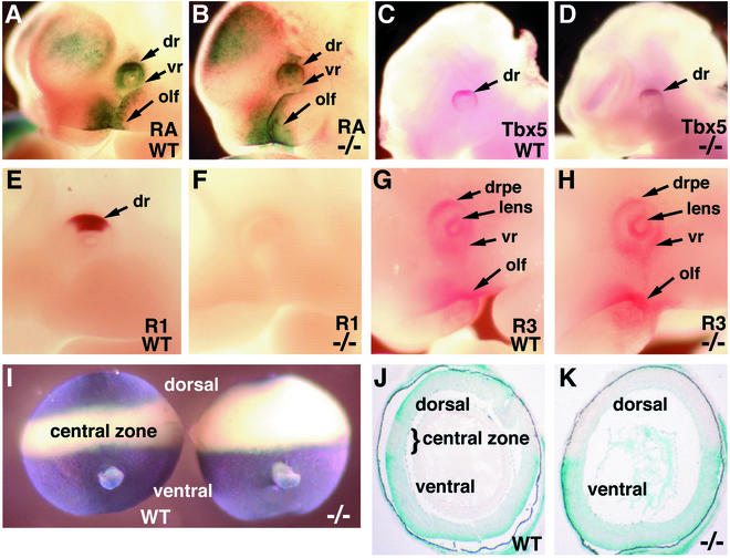
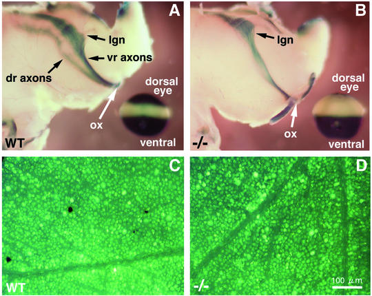
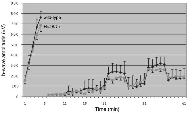
Genetic studies have shown that retinoic acid (RA) signaling is required for mouse retina development, controlled in part by an RA-generating aldehyde dehydrogenase encoded by Aldh1a2 (Raldh2) expressed transiently in the optic vesicles. We examined the function of a related gene, Aldh1a1 (Raldh1), expressed throughout development in the dorsal retina. Raldh1(-/-) mice are viable and exhibit apparently normal retinal morphology despite a complete absence of Raldh1 protein in the dorsal neural retina. RA signaling in the optic cup, detected by using a RARE-lacZ transgene, is not significantly altered in Raldh1(-/-) embryos at embryonic day 10.5, possibly due to normal expression of Aldh1a3 (Raldh3) in dorsal retinal pigment epithelium and ventral neural retina. However, at E16.5 when Raldh3 is expressed ventrally but not dorsally, Raldh1(-/-) embryos lack RARE-lacZ expression in the dorsal retina and its retinocollicular axonal projections, whereas normal RARE-lacZ expression is detected in the ventral retina and its axonal projections. Retrograde labeling of adult Raldh1(-/-) retinal ganglion cells indicated that dorsal retinal axons project to the superior colliculus, and electroretinography revealed no defect of adult visual function, suggesting that dorsal RA signaling is unnecessary for retinal ganglion cell axonal outgrowth. We observed that RA synthesis in liver of Raldh1(-/-) mice was greatly reduced, thus showing that Raldh1 indeed participates in RA synthesis in vivo. Our findings suggest that RA signaling may be necessary only during early stages of retina development and that if RA synthesis is needed in dorsal retina, it is catalyzed by multiple enzymes, including Raldh1.


Figures

FIG. 1.
FIG. 1.
Raldh1 gene targeting. (A) The wild-type Raldh1 gene contains 13 exons, and gene targeting with the replacement vector shown creates a mutant Raldh1 locus in which exon 11 has been deleted. (B) Southern blot showing the genotypes of mice obtained from heterozygous matings. (C) Western blot showing a complete lack of Raldh1 protein detection in Raldh1−/− adult liver (Liv), lung (Lu), and testis (Tes), whereas Raldh1 protein is detected in all three tissues of wild-type mice; as a control, Adh3 protein is detected in both Raldh1−/− and wild-type tissues.
FIG. 2.
FIG. 2.
Analysis of retinal morphology in newborn and adult Raldh1−/− mice. Hematoxylin-eosin staining is shown for retinal sections of wild-type (WT) newborn dorsal retina (A), Raldh1−/− newborn dorsal retina (B), WT newborn ventral retina (C), and Raldh1−/− newborn ventral retina (D). Also, shown is hematoxylin-eosin staining of adult dorsal retina sections for WT (E) and Raldh1−/− (F) mice. Bar, 50 μm (E and F). GCL, ganglion cell layer; INL, inner nuclear layer; ONL, outer nuclear layer.
FIG. 3.
FIG. 3.
Effect of Raldh1−/− genotype on the embryonic dorsal retina. (A and B) RA detection in E10.5 wild-type (WT) and Raldh1−/− embryos carrying the RARE-lacZ transgene. (C and D) Tbx5 mRNA observed at E10.5 by whole-mount in situ hybridization. (E to G) Raldh1 protein (E and F) and Raldh3 protein (G and H), both detected at E10.5 by whole-mount immunohistochemistry. (I) RA detection in E16.5 dissected whole eyes by using the RARE-lacZ transgene (shown from a posterior view with optic nerve observed in the ventral portion). (J and K) Frontal sections through the eyes shown in panel I. dr, dorsal retina; drpe, dorsal RPE; lens, lens vesicle; olf, olfactory region; R1, Raldh1 protein; R3, Raldh3 protein; vr, ventral retina.
FIG. 4.
FIG. 4.
Investigation of retinal ganglion cell axonal projections to the brain in Raldh1−/− mice. (A and B) RARE-lacZ expression in retinal ganglion cell axonal projections into the embryonic brain at E16.5 in wild-type (WT) (A) and Raldh1−/− (B) embryos; in the lower left corner a RARE-lacZ-stained eye from the same embryo is shown for orientation. It should be stressed that when the dorsal and ventral retina axon tracts pass beyond the optic chiasm, they innervate the spatially reversed portions of the brain (ventral-lower and dorsal-upper regions, respectively). (C and D) For adult mice, retrograde labeling of retinal ganglion cells is shown for the WT (C) and Raldh1−/− (D) genotypes. Bar, 100 μm (C and D). dr, dorsal retina; lgn, lateral geniculate nucleus; ox, optic chiasm; vr, ventral retina.
FIG. 5.
FIG. 5.
Visual function assessed by ERG. Female wild-type mice (n = 5) and Raldh1−/− mice (n = 4) at approximately 210 days of age were examined. No statistically significant difference was observed between the two genotypes. Error bars indicate standard deviations.
FIG. 6.
FIG. 6.
Effect of Raldh1 genotype on retinoid levels following a dose of retinol. all-trans-RA and all-trans-retinol were examined by HPLC in liver 2 h after a 50-mg/kg oral dose of all-trans-retinol. Shown are HPLC chromatograms at 355 nm (A to C) and, for each chromatogram, the corresponding spectra of peak 2 showing λmax (A′ to C′). (A) Characteristic retention times for 2-μg standard samples of all-trans-retinol (peak 1) and all-trans-RA (peak 2). (B and C) Wild-type liver (B) and Raldh1−/− liver (C) following retinol treatment (in both cases, 25-μl samples representing 75 mg of liver were loaded). The spectrum of all-trans-RA (peak 2) in the standards (A′) is very similar to the spectra of peak 2 in wild-type liver (B′) and Raldh1−/− liver (C′), thus verifying peak 2 as all-trans-RA in the liver samples. Also, the spectrum for all-trans-retinol (peak 1) in the standards matched the spectra for peak 1 in the liver samples (not shown). AU, absorbance units.

Similar articles

Cited by

References

    1. Ang, H. L., and G. Duester. 1999. Retinoic acid biosynthetic enzyme ALDH1 localizes in a subset of retinoid-dependent tissues during Xenopus development. Dev. Dyn. 215:264-272. - PubMed
    1. Ang, H. L., and G. Duester. 1999. Stimulation of premature retinoic acid synthesis in Xenopus embryos following premature expression of aldehyde dehydrogenase ALDH1. Eur. J. Biochem. 260:227-234. - PubMed
    1. Chapman, D. L., N. Garvey, S. Hancock, M. Alexiou, S. I. Agulnik, J. J. Gibson-Brown, J. Cebra-Thomas, R. J. Bollag, L. M. Silver, and V. E. Papaioannou. 1996. Expression of the T-box family genes, Tbx1-Tbx5, during early mouse development. Dev. Dyn. 206:379-390. - PubMed
    1. Collins, M. D., C. Eckhoff, I. Chahoud, G. Bochert, and H. Nau. 1992. 4-Methylpyrazole partially ameliorated the teratogenicity of retinol and reduced the metabolic formation of all-trans-retinoic acid in the mouse. Arch. Toxicol. 66:652-659. - PubMed
    1. Deltour, L., R. J. Haselbeck, H. L. Ang, and G. Duester. 1997. Localization of class I and class IV alcohol dehydrogenases in mouse testis and epididymis: potential retinol dehydrogenases for endogenous retinoic acid synthesis. Biol. Reprod. 56:102-109. - PubMed

Publication types

MeSH terms