. 2003 Jul;23(13):4663-72.
doi: 10.1128/MCB.23.13.4663-4672.2003.

The Bcr kinase downregulates Ras signaling by phosphorylating AF-6 and binding to its PDZ domain

Affiliations

The Bcr kinase downregulates Ras signaling by phosphorylating AF-6 and binding to its PDZ domain

G Radziwill et al. Mol Cell Biol. 2003 Jul.

Abstract

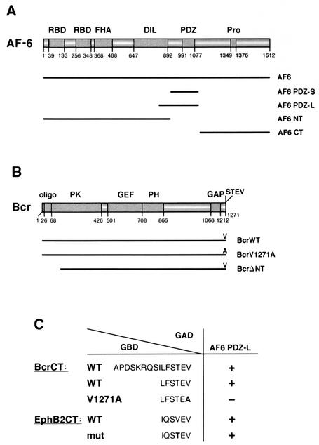
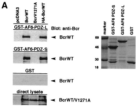
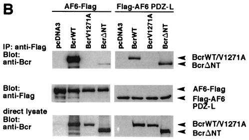
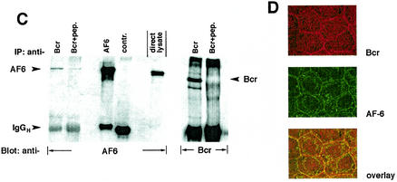
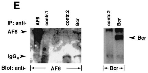
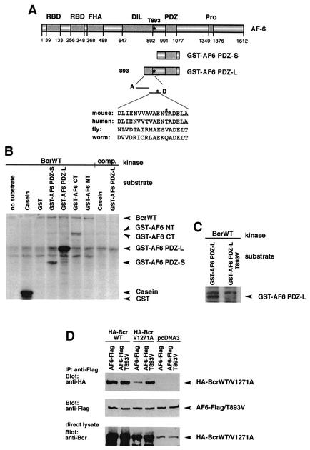
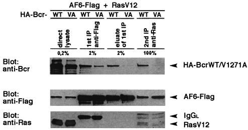
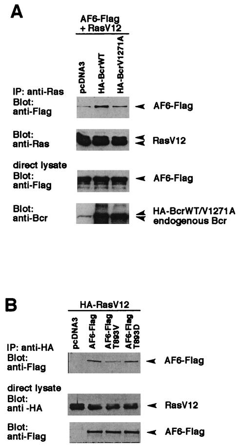
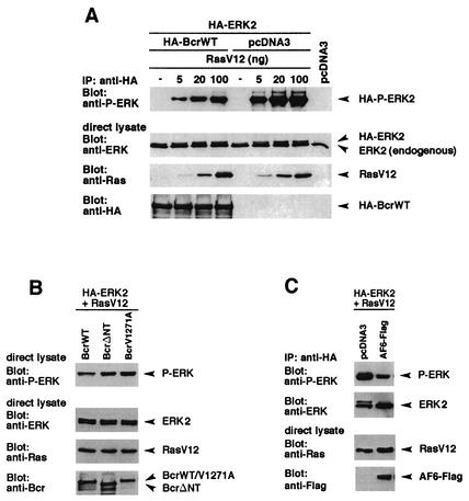
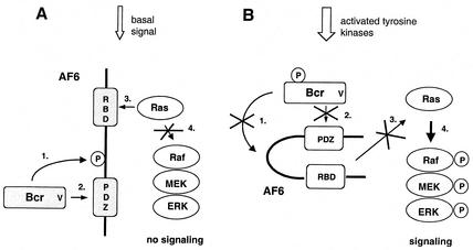
The protein kinase Bcr is a negative regulator of cell proliferation and oncogenic transformation. We identified Bcr as a ligand for the PDZ domain of the cell junction and Ras-interacting protein AF-6. The Bcr kinase phosphorylates AF-6, which subsequently allows efficient binding of Bcr to AF-6, showing that the Bcr kinase is a regulator of the PDZ domain-ligand interaction. Bcr and AF-6 colocalize in epithelial cells at the plasma membrane. In addition, Bcr, AF-6, and Ras form a trimeric complex. Bcr increases the affinity of AF-6 to Ras, and a mutant of AF-6 that lacks a specific phosphorylation site for Bcr shows a reduced binding to Ras. Wild-type Bcr, but not Bcr mutants defective in binding to AF-6, interferes with the Ras-dependent stimulation of the Raf/MEK/ERK pathway. Since AF-6 binds to Bcr via its PDZ domain and to Ras via its Ras-binding domain, we propose that AF-6 functions as a scaffold-like protein that links Bcr and Ras to cellular junctions. We suggest that this trimeric complex is involved in downregulation of Ras-mediated signaling at sites of cell-cell contact to maintain cells in a nonproliferating state.


Figures

FIG. 1.
FIG. 1.
Bcr is a ligand of the PDZ domain of AF-6. (A) Scheme of the domain structure of AF-6. Numbers indicate the amino acid positions. The human AF-6 contains two Ras-binding domains (RBD), a forkhead-associated (FHA) domain, a class V myosin homology region named the DIL domain, a PDZ domain, and a proline-rich region (Pro). Constructs used in this study express full-length AF-6 (AF6), the N terminus of AF-6 corresponding to residues 1 to 914 (AF6 NT), the PDZ domain comprising residues 915 to 1129 (AF6 PDZ-S), and the C terminus of AF-6 containing residues 1130 to 1612 (AF6 CT). AF6 PDZ-L is expressed by a mouse AF-6 cDNA clone coding for the residues 867 to 1145 of mouse AF-6 that correspond to amino acids 850 to 1129 of human AF-6 (4). (B) Scheme of the domain structure of Bcr. Numbers indicate the amino acid positions. Bcr contains an oligomerization sequence (oligo), a serine/threonine protein kinase (PK) domain, a GEF function, a pleckstrin homology (PH) domain, and a GAP domain. At the extreme C terminus, Bcr contains a PDZ domain-binding motif (STEV). Bcr constructs used express full-length Bcr (BcrWT), a mutant in which the C-terminal valine is replaced by alanine (BcrV1271A), and a kinase-defective N-terminal deletion mutant that codes for residues 168 to 1271 (BcrΔNT). Notice that the mutant BcrΔNT still contains the PDZ domain-binding motif. (C) C-terminal sequences of Bcr and EphB2 were tested for their ability to bind to the AF-6 PDZ domain (AF6 PDZ-L) in a Gal4-dependent yeast two-hybrid assay (5). The wild-type sequences contain the C-terminal 15 or 6 residues of Bcr (BcrCT) or EphB2 (EphB2CT) as indicated. The construct BcrCT V1271A contains the C-terminal residue alanine instead of valine, which destroys the putative PDZ domain-binding motif. The construct EphB2CTmut comprises threonine instead of valine at position −2, where the last residue indicates position 0. This construct resembles the C terminus of Bcr and has been shown to bind to the AF-6 PDZ domain (13). All C-terminal sequences were expressed as fusions with the Gal4 DNA-binding domain (GBD), whereas the PDZ domain of AF-6 (AF6 PDZ-L) was expressed as a fusion to the Gal4 activation domain (GAD). Interaction between the C-terminal sequences and the PDZ domain of AF-6 was detected by a β-galactosidase activity assay; interaction is indicated by a plus, no interaction is indicated by a minus (38).
FIG.2.
FIG.2.
Bcr binds to AF-6 in a PDZ domain-dependent manner in vitro and in vivo. (A) HEK293 cells transfected with plasmids coding for BcrWT, BcrV1271A, and HA-BcrWT or empty vector (pcDNA3) were lysed and incubated with GST-AF6 PDZ-L, GST-AF6 PDZ-S, or GST immobilized on GSH-agarose beads as indicated. Proteins bound to GST fusion proteins were fractionated by SDS-PAGE and were immunoblotted with anti-Bcr antibody. As control for the expression of Bcr proteins, direct lysates of 293 cells were immunoblotted with anti-Bcr antibody. The amount of the GST fusion proteins was controlled by Coomassie brilliant blue staining of the SDS-polyacrylamide gel (right panel). (B) HEK293 cells were cotransfected with plasmids coding for AF6-Flag or Flag-AF6 PDZ-L together with Bcr plasmids as indicated. Cell lysates were incubated with anti-Flag antibody (IP). Coprecipitation of Bcr proteins was analyzed by immunoblotting (Blot) with anti-Bcr antibody. Efficiency of immunoprecipitation of Flag-tagged AF6 proteins with anti-Flag antibody and the amount of Bcr protein in direct lysates was monitored by immunoblotting with anti-Flag and anti-Bcr antibody, respectively. (C) Coprecipitation of endogenous AF-6 and Bcr in MDCK cells. The lysate of MDCK cells was incubated with anti-Bcr antibody in the absence or presence of Bcr peptide (+pep.) followed by immunoblotting with anti-AF-6 antibody (lanes 1 and 2). Expression of endogenous AF-6 and Bcr was verified by immunoprecipitation with anti-AF6 and anti-Bcr antibody, respectively, and immunoblotting with the same antibody (lanes 3 and 6) and by analysis of direct lysate with anti-AF-6 antibody (lane 5). A control mouse MAb (anti-cdc2/p34) (lane 4) and the Bcr peptide used as antigen (lane 7) proved the specificity of the antibody reaction. (D) Endogenous Bcr and AF-6 colocalize in MDCK cells. Confluent MDCK cells were double stained with rabbit polyclonal antibody against anti-Bcr and mouse MAb against anti-AF-6. As secondary antibodies rhodamine-conjugated anti-rabbit IgGs and fluorescein-conjugated anti-mouse IgGs were used. (E) Coprecipitation of endogenous AF-6 and Bcr in mouse brain extract. The lysate was treated as indicated and described in (C). Contr. 1, mouse monoclonal anti-cdc2/p34 antibody; Contr. 2, rabbit polyclonal anti-cdk2 antibody.
FIG.2.
FIG.2.
Bcr binds to AF-6 in a PDZ domain-dependent manner in vitro and in vivo. (A) HEK293 cells transfected with plasmids coding for BcrWT, BcrV1271A, and HA-BcrWT or empty vector (pcDNA3) were lysed and incubated with GST-AF6 PDZ-L, GST-AF6 PDZ-S, or GST immobilized on GSH-agarose beads as indicated. Proteins bound to GST fusion proteins were fractionated by SDS-PAGE and were immunoblotted with anti-Bcr antibody. As control for the expression of Bcr proteins, direct lysates of 293 cells were immunoblotted with anti-Bcr antibody. The amount of the GST fusion proteins was controlled by Coomassie brilliant blue staining of the SDS-polyacrylamide gel (right panel). (B) HEK293 cells were cotransfected with plasmids coding for AF6-Flag or Flag-AF6 PDZ-L together with Bcr plasmids as indicated. Cell lysates were incubated with anti-Flag antibody (IP). Coprecipitation of Bcr proteins was analyzed by immunoblotting (Blot) with anti-Bcr antibody. Efficiency of immunoprecipitation of Flag-tagged AF6 proteins with anti-Flag antibody and the amount of Bcr protein in direct lysates was monitored by immunoblotting with anti-Flag and anti-Bcr antibody, respectively. (C) Coprecipitation of endogenous AF-6 and Bcr in MDCK cells. The lysate of MDCK cells was incubated with anti-Bcr antibody in the absence or presence of Bcr peptide (+pep.) followed by immunoblotting with anti-AF-6 antibody (lanes 1 and 2). Expression of endogenous AF-6 and Bcr was verified by immunoprecipitation with anti-AF6 and anti-Bcr antibody, respectively, and immunoblotting with the same antibody (lanes 3 and 6) and by analysis of direct lysate with anti-AF-6 antibody (lane 5). A control mouse MAb (anti-cdc2/p34) (lane 4) and the Bcr peptide used as antigen (lane 7) proved the specificity of the antibody reaction. (D) Endogenous Bcr and AF-6 colocalize in MDCK cells. Confluent MDCK cells were double stained with rabbit polyclonal antibody against anti-Bcr and mouse MAb against anti-AF-6. As secondary antibodies rhodamine-conjugated anti-rabbit IgGs and fluorescein-conjugated anti-mouse IgGs were used. (E) Coprecipitation of endogenous AF-6 and Bcr in mouse brain extract. The lysate was treated as indicated and described in (C). Contr. 1, mouse monoclonal anti-cdc2/p34 antibody; Contr. 2, rabbit polyclonal anti-cdk2 antibody.
FIG. 3.
FIG. 3.
Bcr phosphorylates AF-6 at threonine 893. (A) Schematic representation of AF-6 and its constructs used for the in vitro kinase assay. Peptides A and B represent the sequence unique to GST-AF6 PDZ-L. The sequence of peptide B containing T893 is depicted. This sequence is highly conserved among human and mouse AF-6 but is less conserved in the AF-6 homologs of fly and worm that lack the putative phosphorylation site at the position corresponding to T893. (B) Bcr phosphorylates AF-6. Immunopurified BcrWT prepared from HEK293 cells overexpressing BcrWT was used as kinase. The following GST proteins served as substrates: GST fused to AF6 PDZ-S, AF6 PDZ-L, AF6 CT, and AF6 NT. The specificity of the Bcr kinase activity was checked by using casein and GST alone as positive and negative controls, respectively, and by competition (comp.) of the anti-Bcr antibody with its peptide antigen. The amount of Bcr and of GST fusion proteins was verified by immunoblotting with anti-Bcr and anti-GST antibody (data not shown). (C) Bcr phosphorylates AF-6 at Thr 893. In vitro kinase assays were performed as described for panel B. As substrate, GST-AF6 PDZ-L and GST-AF6 PDZ-L T893V were tested. (D) Thr 893 of AF-6 regulates binding to BcrV1271A. HEK293 cells were transfected with Bcr and AF-6 constructs as indicated. Lysates were incubated with anti-Flag antibody, and coprecipitation of HA-Bcr proteins was analyzed by immunoblotting with anti-HA antibody. Expression of AF-6 and Bcr proteins was controlled by immunoblotting the direct lysate with the appropriate antibody. WT, wild type.
FIG. 4.
FIG. 4.
Bcr, AF-6, and Ras form a trimeric complex. HEK293 cells were cotransfected with BcrWT or BcrV1271A, respectively, together with RasV12 and AF6-Flag. The expression of the proteins was confirmed directly by immunoblot (direct lysate). The first immunoprecipitation (1st IP) was performed with anti-Flag antibody. The Flag complex was eluted from the antibody by incubation with Flag peptide (eluate of 1st IP) and was reprecipitated with anti-Ras antibody (2nd IP). With the exception of the second immunoprecipitation, only small aliquots of the total sample were loaded on the gel as indicated. Bcr, AF-6, and Ras were detected by immunoblotting with appropriate antibodies. IgGL, Ig light chain.
FIG. 5.
FIG. 5.
Bcr modulates the formation of the AF-6/Ras complex. (A) Coexpression of BcrWT increases the complex formation between AF-6 and RasV12. HEK293 cells were transfected with BcrWT or BcrV1271A, respectively, together with RasV12 and AF6-Flag. Cell lysates were incubated with anti-Ras antibody, and coprecipitation of AF6-Flag was detected by immunoblotting with anti-Flag antibody. The amount of precipitated RasV12 and the expression of Bcr and AF-6 were checked by immunoblotting with the appropriate antibodies as indicated. (B) Prevention of phosphorylation of AF-6 by Bcr reduces binding of AF-6 to Ras. HA-RasV12 was coexpressed with AF6-Flag or AF-6 mutant proteins, AF6 T893V and AF6 T893D, that carry a single amino acid exchange at position 893 from Thr to Val or Asp, respectively. Cell lysates were incubated with anti-HA antibody for immunoprecipitation of HA-RasV12, and coprecipitation of AF6-Flag proteins was detected by immunoblotting with anti-Flag antibody. Expression of HA-RasV12 and AF6-Flag proteins was monitored by immunoblotting with the appropriate antibodies as indicated. IP, immunoprecipitation.
FIG. 6.
FIG. 6.
Bcr interferes with Ras-mediated ERK2 activation in a PDZ ligand-dependent manner. (A) HEK293 cells plated on 6-cm-diameter dishes were cotransfected with plasmids coding for HA-BcrWT or empty vector (4 μg) together with HA-ERK2 (1 μg) and increasing amounts of RasV12 as indicated. Cell lysates were incubated with anti-HA antibody, and the activity of immunoprecipitated HA-ERK2 was determined by immunoblotting with anti-phospho-ERK2 (anti-P-ERK) antibody. Expression of HA-BcrWT, HA-ERK2, and RasV12 was verified by immunoblotting with the appropriate antibodies as indicated. (B) HEK293 cells, which expressed BcrWT, BcrΔNT, or BcrV1271A together with HA-ERK2 and RasV12 (20 ng) were analyzed for the amount of phosphorylated, activated ERK2 as described for panel A. Expression of proteins was controlled by immunoblotting with the appropriate antibodies as indicated. (C) AF-6 can reduce Ras-mediated ERK2 activation. AF-6 and control vector were transfected in HEK293 cells together with RasV12 and HA-ERK and were analyzed as described for panel A. IP, immunoprecipitation.
FIG. 7.
FIG. 7.
Proposed model depicting the effect of Bcr on Ras-dependent stimulation of ERK via AF-6. (A) In quiescent cells the constitutively active Bcr phosphorylates AF-6 (step 1), which leads to the interaction of the PDZ domain of AF-6 with the PDZ-binding motif of Bcr (step 2). This interaction increases the affinity of AF-6 for Ras via the Ras binding domain (RBD) (step 3) and prevents binding of Raf to Ras (step 4). Under these conditions the protein kinase cascade composed of Raf, MEK, and ERK is not activated. (B) Phosphorylation of Bcr on tyrosine residues inactivates its protein kinase activity. Therefore, Bcr cannot phosphorylate (step 1) and cannot bind to AF-6 (step 2). Thus, AF-6 does not compete with Raf for Ras (step 3) and does not interfere with the Ras-dependent activation of the protein kinase cascade (step 4). P represents phosphorylation of proteins.

Similar articles

Cited by

References

    1. Boettner, B., E. Govek, J. Cross, and L. van Aelst. 2000. The junctional multidomain protein AF-6 is a binding partner of the Rap1A GTPase and associates with the actin cytoskeletal regulator profilin. Proc. Natl. Acad. Sci. USA 97:9064-9069. - PMC - PubMed
    1. Boguski, M. S., and F. McCormick. 1993. Proteins regulating Ras and its relatives. Nature 366:643-654. - PubMed
    1. Braselmann, S., and F. McCormick. 1995. Bcr and Raf form a complex in vivo via 14-3-3 proteins. EMBO J. 14:4839-4848. - PMC - PubMed
    1. Buchert, M., S. Schneider, V. Meskenaite, M. T. Adams, E. Canaani, T. Baechi, K. Moelling, and C. M. Hovens. 1999. The junction-associated protein AF-6 interacts and clusters with specific Eph receptor tyrosine kinases at specialized sites of cell-cell contact in the brain. J. Cell Biol. 144:361-371. - PMC - PubMed
    1. Durfee, T., K. Becherer, P. L. Chen, S. H. Yeh, Y. Yang, A. E. Kilburn, W. H. Lee, and S. J. Elledge. 1993. The retinoblastoma protein associates with the protein phosphatase type 1 catalytic subunit. Genes Dev. 7:555-569. - PubMed

Publication types

MeSH terms