. 2003 Jun 24;100(13):7889-94.
doi: 10.1073/pnas.1330742100. Epub 2003 Jun 13.

Crystal structure of the SOS cell division inhibitor SulA and in complex with FtsZ

Affiliations

Crystal structure of the SOS cell division inhibitor SulA and in complex with FtsZ

Suzanne C Cordell et al. Proc Natl Acad Sci U S A. .

Abstract

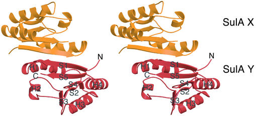
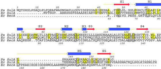
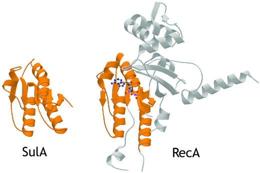
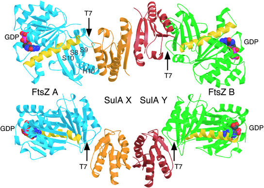
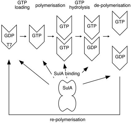
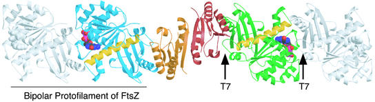
SulA halts cell division in Escherichia coli by binding to the major component of the division machinery FtsZ. We have solved the crystal structure of SulA alone and in complex with FtsZ from Pseudomonas aeruginosa. SulA is expressed when the SOS response is induced. This is a mechanism to inhibit cell division and repair DNA in the event of DNA damage. FtsZ is a tubulin-like protein that forms polymers, with the active-site GTPase split across two monomers. One monomer provides the GTP-binding site and the other, through its T7 loop nucleotide hydrolysis. Our structures show that SulA is a dimer, and that SulA inhibits cell division neither by binding the nucleotide-binding site nor by inducing conformational changes in FtsZ. Instead, SulA binds the T7 loop surface of FtsZ, opposite the nucleotide-binding site, blocking polymer formation. These findings explain why GTP hydrolysis and polymer turnover are required for SulA inhibition.


Figures

Fig. 1.
Fig. 1.
P. aeruginosa SulA has a fold similar to the nucleotide-binding core of RecA and forms a dimer. Stereo ribbon drawing of the SulA dimer with one monomer shown in orange and the other in red.
Fig. 2.
Fig. 2.
Structure-based sequence alignment [P. aeruginosa SulA homolog, Swiss-Prot Q9HZJ8, PDB ID code 1OFT, E. coli SulA, Swiss-Prot SULA_ECOLI, E. coli RecA, Swiss-Prot RECA_ECOLI, PDB ID code 2REB (33)]. Pa and Ec SulA have been aligned purely on the basis of their sequences. The overall identity between Pa and Ec SulA is 32%. The structures of Pa SulA and Ec RecA have been superimposed, and the resulting sequence alignment is shown. Only the N-terminal domain of RecA superimposes with SulA.
Fig. 3.
Fig. 3.
Structural similarity of the SulA monomer (PDB ID code 1OFT) and the N-terminal domain of E. coli RecA (PDB ID code 2REB; ref. 33). The structures have been aligned with an rms deviation of 2.4 Å over 112 Cα atoms (of 119 for SulA).
Fig. 4.
Fig. 4.
Crystal structure (2.1 Å) of the SulAΔ35:FtsZ complex (PDB ID code 1OFU). The SulA dimer is sandwiched between two FtsZ monomers via the T7 protofilament interface, leaving the GTP-binding regions exposed. Note how the FtsZ molecules are rotated exactly 180° relative to each other in the complex. The SulA dimer is in orange and red as in Fig. 1. The FtsZ molecules are shown in blue and green. The yellow helix is H7 and marks the transition from the N- to the C-terminal domain (8). GDP is shown in space-filling representation. The two views are rotated by 90° about x.
Fig. 5.
Fig. 5.
Schematic drawing of the FtsZ polymerization/depolymerization cycle and SulA inhibition. FtsZ can polymerize only when in the GTP state. Once polymerized, FtsZ will hydrolyze GTP to GDP, because polymerization allows FtsZ to form complete active sites as they are split across two monomers. Depolymerization occurs some time after GTP hydrolysis. SulA, as shown in this study, will bind to FtsZ containing GTP and GDP. Because one of the two protofilament contacts is still available in the SulA:FtsZ complex structure, it is possible to bind the ends of protofilaments. The complex between SulA and FtsZ is very tight, and the FtsZ polymerization cycle means that each FtsZ molecule will at some point expose a free T7 interface. SulA will bind to this interface and remove it from the pool of available T7 interfaces for polymerization. Because SulA is produced in large amounts during SOS response, eventually all FtsZ will be monomeric and polymerization inhibited. Lon protease is needed to free FtsZ from SulA.
Fig. 6.
Fig. 6.
Model showing how SulA could crosslink the ends of FtsZ protofilaments. The complex is as in Fig. 4. FtsZ monomers, shown in gray, could theoretically bind to each and of the complex in a protofilament-like structure where the T7 loop from one monomer contacts the GTP-binding site from the previous monomer (17).

References

    1. Fernandez de Henestrosa, A. R., Ogi, T., Aoyagi, S., Chafin, D., Hayes, J. J., Ohmori, H. & Woodgate, R. (2000) Mol. Microbiol. 35, 1560-1572. - PubMed
    1. Walker, G. C. (1995) Trends Biochem. Sci. 20, 416-420. - PubMed
    1. Husiman, O. & Dari, R. (1981) Nature 290, 797-799. - PubMed
    1. Husiman, O., Dari, R. & Gottesman, S. (1984) Proc. Natl. Acad. Sci. USA 81, 4490-4494. - PMC - PubMed
    1. Mizusawa, S. & Gottesman, S. (1983) Proc. Natl. Acad. Sci. USA 80, 358-362. - PMC - PubMed

LinkOut - more resources