. 2003 Jul 8;100(14):8430-5.
doi: 10.1073/pnas.0932636100. Epub 2003 Jun 13.

Transforming growth factor beta signaling impairs Neu-induced mammary tumorigenesis while promoting pulmonary metastasis

Affiliations

Transforming growth factor beta signaling impairs Neu-induced mammary tumorigenesis while promoting pulmonary metastasis

Peter M Siegel et al. Proc Natl Acad Sci U S A. .

Abstract

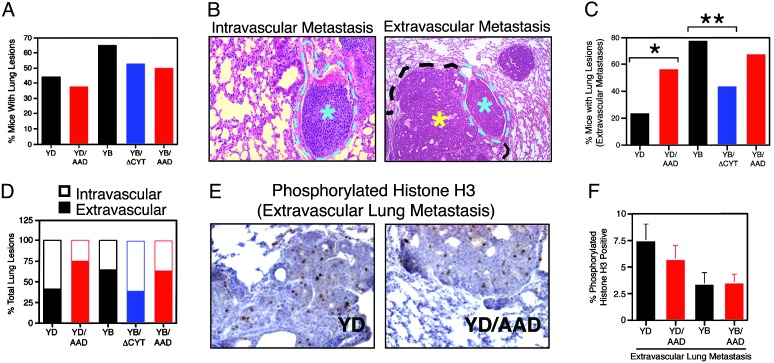
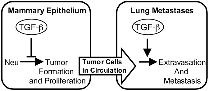
The influence of transforming growth factor beta (TGF-beta) signaling on Neu-induced mammary tumorigenesis and metastasis was examined with transgenic mouse models. We generated mice expressing an activated TGF-beta type I receptor or dominant negative TGF-beta type II receptor under control of the mouse mammary tumor virus promoter. When crossed with mice expressing activated forms of the Neu receptor tyrosine kinase that selectively couple to the Grb2 or Shc signaling pathways the activated type I receptor increased the latency of mammary tumor formation but also enhanced the frequency of extravascular lung metastasis. Conversely, expression of the dominant negative type II receptor decreased the latency of Neu-induced mammary tumor formation while significantly reducing the incidence of extravascular lung metastases. These observations argue that TGF-beta can promote the formation of lung metastases while impairing Neu-induced tumor growth and suggest that extravasation of breast cancer cells from pulmonary vessels is a point of action of TGF-beta in the metastatic process.


Figures

Fig. 1.
Fig. 1.
Decreased proliferation and enhanced cell death diminish mammary epithelial cell density in TβRI(AAD) transgenic mice. Shown are schematic diagrams of the MMTV/TβRI(AAD) transgene indicating the position of activating mutations (*) within the GS domain (A) and the MMTV/TβRII(ΔCyt) transgene highlighting insertion of a stop codon downstream of the transmembrane domain (B), depicted by bold and underlined type. (C) Whole-mount and histological analyses were conducted on control (FVB/N) and transgenic (AAD) mice from day-15 pregnant (D15P) and lactating mammary glands (L.M.GL.). (Left) Scale bars represent 0.5 cm. (Right) Arrows indicate the region magnified in the Inset. Representative sections illustrating the appearance of BrdUrd-positive cells in day-12 pregnant (D12P) mice (D) and apoptotic cells (arrows) in day-15 pregnant (D15P) mammary glands of control (FVB/N) and transgenic (AAD) mice (E). Scale bars represent 100 μm. The percentage of BrdUrd and terminal deoxynucleotidyltransferase-mediated dUTP nick end labeling-positive cells in FVB/N (open bars) and AAD (filled bars) mammary glands is shown during different stages of mammary gland development (D and E Bottom).
Fig. 2.
Fig. 2.
TGF-β signaling delays Neu-induced mammary tumorigenesis. (A) The Neu receptors harbor an oncogenic cysteine deletion within the extracellular domain (star) and tyrosine to phenylalanine substitutions of autophosphorylation sites within the cytoplasmic tail. A single phenylalanine was reverted back to a tyrosine to restore either the Shc (YD) or Grb2 (YB) binding site. (B) Kinetics of mammary tumor formation in Neu(YD) versus YD/AAD mice (P = 0.0001, Student's t test). (C) Mammary tumor onset in Neu(YB) versus YB/AAD (P = 0.0002, Student's t test) and Neu(YB) versus YB/ΔCyt (P = 0.04, Student's t test) transgenic mice. Age of onset is the time that a palpable mammary tumor first appears. T50 denotes the age at which 50% of the mice first possess a tumor, and n is the number of mice examined.
Fig. 3.
Fig. 3.
Neu(YD) and Neu(YB) mammary tumor growth is inhibited by TGF-β. (A) Immunostaining of phosphorylated histone H3 reveals a significant reduction in the percentage of mitotic cells in YD/AAD versus Neu(YD) (*, P = 0.04, Student's t test) and YB/AAD versus Neu(YB) (**, P = 0.01, Student's t test) mammary tumors. (B) Primary mammary tumor cultures from four individual Neu(YD) (red) and Neu(YB) (blue) mice were treated with increasing concentrations of TGF-β in the presence of 125I-deoxyuridine, and the percentage incorporation into DNA relative to control cells without TGF-β stimulation is shown (data are representative of two independent experiments performed with triplicate cultures). Primary mammary epithelial cells isolated from mid-pregnant, nontransgenic mammary glands were included as a control (FVB/N, black). In each case, standard deviations did not exceed 18% of the average value.
Fig. 4.
Fig. 4.
TGF-β signaling enhances the formation of extravascular lung metastases. (A) Percentage of mice with lung lesions; Neu(YD), n = 27; YD/AAD, n = 24; Neu(YB), n = 20; YB/ΔCyt, n = 15; and YB/AAD, n = 12. (B) Lesions were classified as intravascular (light blue asterisk) if they remained confined within a blood vessel (outlined by light blue dotted line) or extravascular (yellow asterisk) if the tumor cells had breached the vessel wall and were growing into the lung parenchyma. (C) The percentage of mice harboring lung lesions that contained extravascular metastases: Neu(YD) versus YD/AAD (*, mid P = 0.05, Fisher's exact test) and Neu(YB) versus YB/ΔCyt (**, mid P = 0.004; Fisher's exact test). (D) The percentage of total lung lesions was segregated into intravascular or extravascular metastases by histological examination (a minimum of 80 lung lesions was scored from each genotype). (E) Representative sections illustrating phosphorylated histone H3-positive cells in Neu(YD) versus YD/AAD extravasated lung lesions. (F) Quantitation of the percentage proliferating cells in extravascular lung lesions from the indicated genotypes.
Fig. 5.
Fig. 5.
TGF-β enhances lung metastases despite suppressive effects on primary tumor formation. The onset of Neu-induced mammary tumors is delayed by expression of an activated TβRI and accelerated by a dominant negative TβRII. However, activation of TGF-β signaling enhances extravasation of Neu-induced mammary tumor cells into lung tissue, which can be impaired by inhibition of the TGF-β pathway.

Comment in

Similar articles

Cited by

References

    1. Massague, J., Blain, S. W. & Lo, R. S. (2000) Cell 103 295-309. - PubMed
    1. Gold, L. I. (1999) Crit. Rev. Oncog. 10 303-360. - PubMed
    1. Oft, M., Peli, J., Rudaz, C., Schwarz, H., Beug, H. & Reichmann, E. (1996) Genes Dev. 10 2462-2477. - PubMed
    1. Portella, G., Cumming, S. A., Liddell, J., Cui, W., Ireland, H., Akhurst, R. J. & Balmain, A. (1998) Cell Growth Differ. 9 393-404. - PubMed
    1. Gorsch, S. M., Memoli, V. A., Stukel, T. A., Gold, L. I. & Arrick, B. A. (1992) Cancer Res. 52 6949-6952. - PubMed

Publication types

MeSH terms

Substances