. 2003 Jul 3;424(6944):76-81.
doi: 10.1038/nature01761. Epub 2003 Jun 15.

Melanopsin and rod-cone photoreceptive systems account for all major accessory visual functions in mice

Affiliations

Melanopsin and rod-cone photoreceptive systems account for all major accessory visual functions in mice

S Hattar et al. Nature. .

Abstract

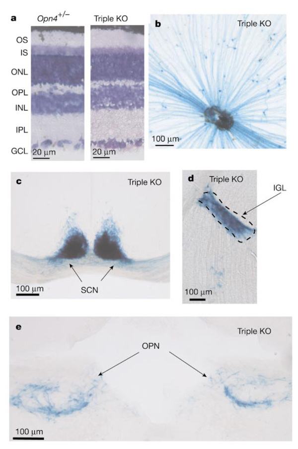
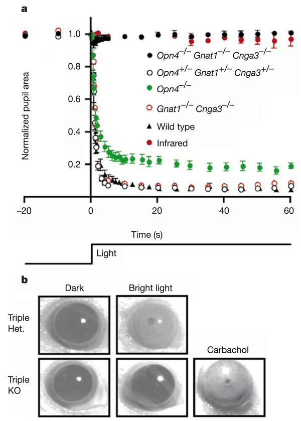
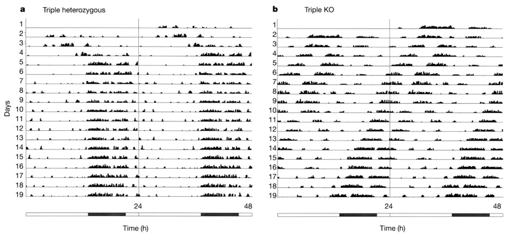
In the mammalian retina, besides the conventional rod-cone system, a melanopsin-associated photoreceptive system exists that conveys photic information for accessory visual functions such as pupillary light reflex and circadian photo-entrainment. On ablation of the melanopsin gene, retinal ganglion cells that normally express melanopsin are no longer intrinsically photosensitive. Furthermore, pupil reflex, light-induced phase delays of the circadian clock and period lengthening of the circadian rhythm in constant light are all partially impaired. Here, we investigated whether additional photoreceptive systems participate in these responses. Using mice lacking rods and cones, we measured the action spectrum for phase-shifting the circadian rhythm of locomotor behaviour. This spectrum matches that for the pupillary light reflex in mice of the same genotype, and that for the intrinsic photosensitivity of the melanopsin-expressing retinal ganglion cells. We have also generated mice lacking melanopsin coupled with disabled rod and cone phototransduction mechanisms. These animals have an intact retina but fail to show any significant pupil reflex, to entrain to light/dark cycles, and to show any masking response to light. Thus, the rod-cone and melanopsin systems together seem to provide all of the photic input for these accessory visual functions.


Figures

Figure 1
Figure 1
Irradiance–response relations (a) and action spectrum (b) for circadian phase shifting in rd/rd cl mice by monochromatic light between 420 nm and 580 nm, assayed by wheel-running (n = 4–7 animals per irradiance at each wavelength). The derived action spectrum in b for circadian photo-entrainment is best fitted (R2 = 0.976) by the absorption spectrum of a vitamin A1-based photopigment with λmax = 481 nm (continuous curve).
Figure 2
Figure 2
Normal retinal morphology and presence and central connectivity of melanopsin-expressing RGCs in triple-knockout (Opn4−/− Gnat1−/− Cnga3−/−) mice. a, Retinal cross-sections from Opn4+/− and triple-knockout (KO) mice. Giemsa stain shows the various layers. Both are similar in morphology and thickness to wild type. GCL, ganglion cell layer; INL, inner nuclear layer; IPL, inner plexiform layer; IS, inner segment layer; ONL, outer nuclear layer; OPL, outer plexiform layer; OS, outer segment layer. b, Flat-mount view of a triple-knockout retina stained with X-gal (blue). ce, Coronal sections of triple-knockout mouse brain showing the normal innervations of the SCN, IGL and OPN by X-gal-labelled axons. Dorsal side is up in each case.
Figure 3
Figure 3
Disabling of rods, cones and melanopsin-positive RGCs essentially eliminates the pupillary light reflex. a, Pupil area (mean ± s.e.m.) as a percentage of dark-adapted aperture area (recorded just before light exposure, time 0 on graph) for Opn4−/− Gnat1−/− Cnga3−/− (n = 6), Opn4+/− Gnat1+/− Cnga3+/− (n = 4), Opn4−/− (n = 8; data reproduced from ref. 8) and Gnat1−/− Cnga3−/− (n = 1) mice over the course of 1 min when exposed to 480-nm light at 86–140 µW cm−2. The wild-type data are from ref. . For comparison, the pupil size of triple-knockout mice exposed to an infrared light pulse (n = 6) is also shown. On close scrutiny, a small, transient pupil constriction was detected in two out of six triple-knockout mice exposed to the intense 480-nm stimulus. The averaged 480-nm data shown for the triple knockouts are from mice dark-adapted for 3 days before light exposure, but are representative of similar recordings from the same group after 1-h dark adaptation. Neither the amplitude nor the frequency of occurrence of the residual pupillary response in the triple-knockout animals were increased with bright white-light stimulus (30 mW cm−2), or with monochromatic stimuli ranging from 360 to 660 nm (70 µW cm−2 at 360 nm; 19 µW cm−2 at 420 nm; 86 µW cm−2 at 480 nm; 100 µW cm−2 at 550 nm; 68 µW cm−2 at 660 nm). Nor did these parameters show any significant change when tested repeatedly over a 24-h period. b, Application of 100-mM carbachol resulted in a complete pupil constriction in triple-knockout mice.
Figure 4
Figure 4
Actograms of wheel running for mice under a 16/8-h light/dark cycle, double-plotted on a 24-h timescale. The illumination was approximately 800 lx white light. The numbered lines represent successive days. Activity levels (in total number of revolutions in 10-min bins) are given in 15 quantiles, with the first being 1–55 revolutions, the second 56–110, and so on. The bar below the actograms indicates light (white) and dark (black) periods. a, Triple heterozygote (Opn4+/− Gnat1+/− Cnga3+/−). The locomotor activity had a cycle very close to 24 h and was phase-locked to darkness, showing photo-entrainment. b, Triple-knockout (Opn4−/− Gnat1−/− Cnga3−/−). The animal free-ran with a period of less than 24 h, showing lack of photo-entrainment.
Figure 5
Figure 5
Actograms of wheel running plotted on a 7-h timescale. The mice were subjected to an ultradian 3.5/3.5-h light/dark cycle (see text). Illumination was approximately 800 lx white light. The numbered lines represent successive 7-h cycles. The activity quantiles and time bins are the same as in Fig. 4 a, b, Two triple-heterozygous mice (Opn4+/− Gnat1+/− Cnga3+/−) showing a negative and positive masking effect, respectively. The percentage of total revolutions in darkness was 99.5% and 32.7%, respectively. b, Two triple-knockout mice (Opn4−/− Gnat1−/− Cnga3−/−). The percentage of total revolutions in darkness was 40.8% and 44.7%, respectively, not far from the 50% expected from randomness; that is, no masking effect of light.

Similar articles

Cited by

  • Autonomic control of the eye.
    McDougal DH, Gamlin PD. McDougal DH, et al. Compr Physiol. 2015 Jan;5(1):439-73. doi: 10.1002/cphy.c140014. Compr Physiol. 2015. PMID: 25589275 Free PMC article. Review.
  • Classical Photoreceptors Are Primarily Responsible for the Pupillary Light Reflex in Mouse.
    Jain V, Srivastava I, Palchaudhuri S, Goel M, Sinha-Mahapatra SK, Dhingra NK. Jain V, et al. PLoS One. 2016 Jun 13;11(6):e0157226. doi: 10.1371/journal.pone.0157226. eCollection 2016. PLoS One. 2016. PMID: 27295136 Free PMC article.
  • Migraine photophobia originating in cone-driven retinal pathways.
    Noseda R, Bernstein CA, Nir RR, Lee AJ, Fulton AB, Bertisch SM, Hovaguimian A, Cestari DM, Saavedra-Walker R, Borsook D, Doran BL, Buettner C, Burstein R. Noseda R, et al. Brain. 2016 Jul;139(Pt 7):1971-86. doi: 10.1093/brain/aww119. Epub 2016 May 17. Brain. 2016. PMID: 27190022 Free PMC article.
  • Melanopsin is highly resistant to light and chemical bleaching in vivo.
    Sexton TJ, Golczak M, Palczewski K, Van Gelder RN. Sexton TJ, et al. J Biol Chem. 2012 Jun 15;287(25):20888-97. doi: 10.1074/jbc.M111.325969. Epub 2012 Apr 30. J Biol Chem. 2012. PMID: 22547062 Free PMC article.
  • Genome of the Chinese tree shrew.
    Fan Y, Huang ZY, Cao CC, Chen CS, Chen YX, Fan DD, He J, Hou HL, Hu L, Hu XT, Jiang XT, Lai R, Lang YS, Liang B, Liao SG, Mu D, Ma YY, Niu YY, Sun XQ, Xia JQ, Xiao J, Xiong ZQ, Xu L, Yang L, Zhang Y, Zhao W, Zhao XD, Zheng YT, Zhou JM, Zhu YB, Zhang GJ, Wang J, Yao YG. Fan Y, et al. Nat Commun. 2013;4:1426. doi: 10.1038/ncomms2416. Nat Commun. 2013. PMID: 23385571

References

    1. Provencio I, Jiang G, DeGrip WJ, Hayes WP, Rollag MD. Melanopsin: an opsin in melanophores, brain, and eye. Proc. Natl Acad. Sci. USA. 1998;95:340–345. - PMC - PubMed
    1. Provencio I, et al. A novel human opsin in the inner retina. J. Neurosci. 2000;20:600–605. - PMC - PubMed
    1. Gooley JJ, Lu J, Chou TC, Scammell TE, Saper CB. Melanopsin in cells of origin of the retinohypothalamic tract. Nature Neurosci. 2001;4:1165. - PubMed
    1. Hannibal J, Hindersson P, Knudsen SM, Georg B, Fahrenkrug J. The photopigment melanopsin is exclusively present in pituitary adenylate cyclase-activating polypeptide-containing retinal ganglion cells of the retinohypothalamic tract. J. Neurosci. 2002;22:RC191. - PMC - PubMed
    1. Provencio I, Rollag MD, Castrucci AM. Photoreceptive net in the mammalian retina. Nature. 2002;415:493. - PubMed

Publication types

MeSH terms