. 2003 Jun 16:3:12.
doi: 10.1186/1471-2180-3-12.

Characterisation of methionine adenosyltransferase from Mycobacterium smegmatis and M. tuberculosis

Affiliations

Characterisation of methionine adenosyltransferase from Mycobacterium smegmatis and M. tuberculosis

Bradley J Berger et al. BMC Microbiol. .

Abstract

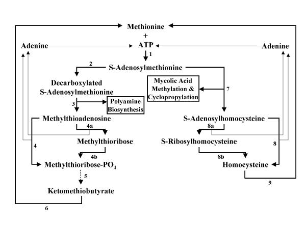
Background: Tuberculosis remains a serious world-wide health threat which requires the characterisation of novel drug targets for the development of future antimycobacterials. One of the key obstacles in the definition of new targets is the large variety of metabolic alterations that occur between cells in the active growth and chronic/dormant phases of tuberculosis. The ideal biochemical target should be active in both growth phases. Methionine adenosyltransferase, which catalyses the formation of S-adenosylmethionine from methionine and ATP, is involved in polyamine biosynthesis during active growth and is also required for the methylation and cyclopropylation of mycolipids necessary for survival in the chronic phase.

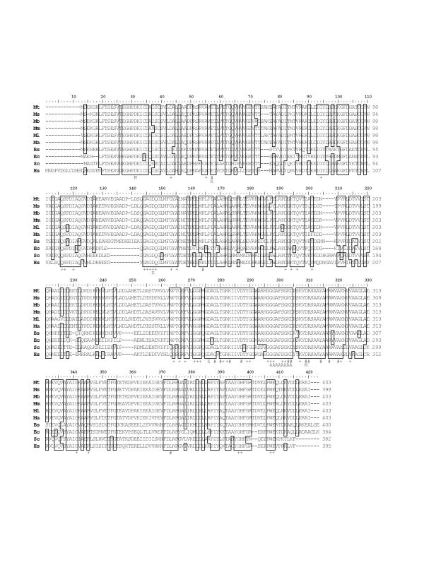
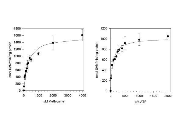
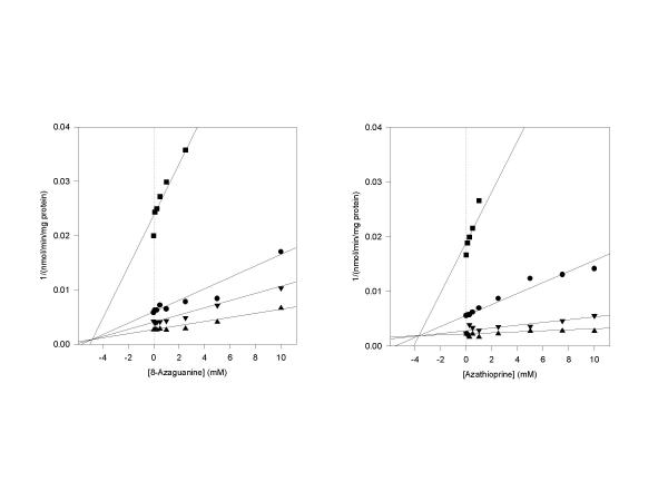
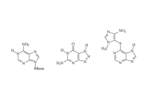
Results: The gene encoding methionine adenosyltransferase has been cloned from Mycobacterium tuberculosis and the model organism M. smegmatis. Both enzymes retained all amino acids known to be involved in catalysing the reaction. While the M. smegmatis enzyme could be functionally expressed, the M. tuberculosis homologue was insoluble and inactive under a large variety of expression conditions. For the M. smegmatis enzyme, the Vmax for S-adenosylmethionine formation was 1.30 micromol/min/mg protein and the Km for methionine and ATP was 288 microM and 76 microM respectively. In addition, the enzyme was competitively inhibited by 8-azaguanine and azathioprine with a Ki of 4.7 mM and 3.7 mM respectively. Azathioprine inhibited the in vitro growth of M. smegmatis with a minimal inhibitory concentration (MIC) of 500 microM, while the MIC for 8-azaguanine was >1.0 mM.

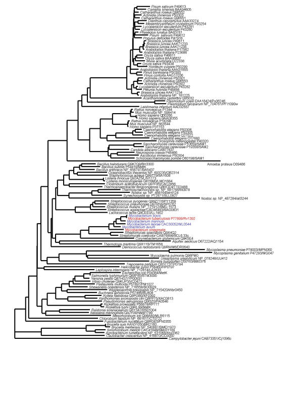
Conclusion: The methionine adenosyltransferase from both organisms had a primary structure very similar those previously characterised in other prokaryotic and eukaryotic organisms. The kinetic properties of the M. smegmatis enzyme were also similar to known prokaryotic methionine adenosyltransferases. Inhibition of the enzyme by 8-azaguanine and azathioprine provides a starting point for the synthesis of higher affinity purine-based inhibitors.


Figures

Figure 1
Figure 1
S-Adenosylmethionine as a common biochemical substrate for the rapid and chronic growth stages of M. tuberculosis. The pathways of S-adenosylmethionine usage and the potential recycling routes of methionine and ATP are shown. The enzymes which catalyse the reactions are: 1 methionine adenosyltransferase, 2 S-adenosylmethionine decarboxylase, 3 spermidine/spermine aminopropyltransferase, 4 methylthioadenosine phosphorylase, 4a methylthioadenosine nucleosidase, 4b methylthioribose kinase, 5 four steps not shown, 6 aminotransferase, 7 mycolic acid methyltransferases, 8 S-adenosylhomocysteine hydrolase, 8a S-adenosylhomocysteine nucleosidase, 8b S-ribosylhomocysteine hydrolase, and 9 methionine synthetase. It has not yet been determined in M. tuberculosis whether enzyme 4 or 4a/4b, and 8 or 8a/8b catalyses the recycling of methionine. The exact aminotransferase catalysing step 6 has also not been elucidated.
Figure 2
Figure 2
Phylogenetic relationship of methionine adenosyltransferase sequences. The enzyme sequences are labelled with Entrez accession numbers and, in the case of microbial genome data, with the protein identifier from the genome project. All sequences were aligned with the Clustal algorithm and used for tree construction using the neighbor-joining method. The sequences from M. tuberculosis and M. smegmatis are in red, while the other mycobacterial sequences are in blue.
Figure 3
Figure 3
Alignment of selected methionine adenosyltransferase sequences. The following sequences were aligned with the Clustal algorithm: Mt, M. tuberculosis; Ms, M. smegmatis; Mb, M. bovis; Mm, M. marinum; Ml, M. leprae; Ma, M. avium; Bs, Bacillus subtilis [51]; Ec, Escherichia coli MetK [52], Sc, Saccharomyces cerevisiae SAM1 [53]; Hs, Homo sapiens MAT1 [54]. Residues conserved by 75% of these sequences are boxed. The annotation below refers to 100% (#) or 98% (+) conservation of residues by the 117 sequences in Figure 2. Residues marked with M are the putative Mg2+ binding sites, K the putative K+ binding sites, A the ATP-binding residues of the P-loop, and X the residues that interact with the methionine substrate.
Figure 4
Figure 4
Kinetic characterisation of M. smegmatis methionine adenosyltransferase. The enzyme was incubated with 0 – 4.0 mM substrate and 10 mM cosubstrate as described in the Methods section. The production of SAM was measured by HPLC, and the resulting data fitted to the Michaelis-Menton equation.
Figure 5
Figure 5
Inhibition of methionine adenosyltransferase by 8-azaguanine and azathioprine. The M. smegmatis enzyme was incubated with 0–10 mM inhibitor, 10 mM methionine, and 0.5 (squares), 1.0 (circles), 2.0 (inverted triangles), or 3.0 (triangles) mM ATP as described in the Methods section. The data is shown as Dixon plots.
Figure 6
Figure 6
The structures of 8-azaguanine and azathioprine. From right to left: the adenine portion of ATP, 8-azaguanine, and azathioprine.

References

    1. Dye C, Scheele S, Dolin P, Pathania V, Raviglione MC. Consensus statement. Global burden of tuberculosis: estimated incidence, prevalence, and mortality by country. WHO Global Surveillance and Monitoring Project. JAMA. 1999;282:677–86. doi: 10.1001/jama.282.7.677. - DOI - PubMed
    1. Besra GS, Chatterjee D. Lipids and carbohydrates of Mycobacterium tuberculosis. In: Bloom BR, editor. Tuberculosis. Pathogenesis, protection, and control. Washington, ASM Press; 1994. pp. 285–306.
    1. Parrish NM, Dick JD, Bishai WR. Mechanisms of latency in Mycobacterium tuberculosis. Tr Microbiol. 1998;6:107–12. doi: 10.1016/S0966-842X(98)01216-5. - DOI - PubMed
    1. McKinney JD, Honer zu Bentrup K, Munoz-Elias EJ, Miczak A, Chen B, Chan WT, Swenson D, Sacchettini JC, Jacobs WR, Jr, Russell DG. Persistence of Mycobacterium tuberculosis in macrophages and mice requires the glyoxylate shunt enzyme isocitrate lyase. Nature. 2000;406:735–8. doi: 10.1038/35021074. - DOI - PubMed
    1. Marton LJ, Pegg AE. Polyamines as targets for therapeutic intervention. Ann Rev Pharmacol Toxicol. 1995;35:55–91. doi: 10.1146/annurev.pharmtox.35.1.55. - DOI - PubMed

Publication types

MeSH terms

LinkOut - more resources