. 2003 Jun 23;161(6):1143-50.
doi: 10.1083/jcb.200210069. Epub 2003 Jun 16.

ADP ribosylation factor 6 is activated and controls membrane delivery during phagocytosis in macrophages

Affiliations

ADP ribosylation factor 6 is activated and controls membrane delivery during phagocytosis in macrophages

Florence Niedergang et al. J Cell Biol. .

Abstract

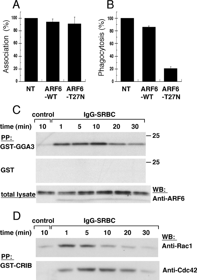
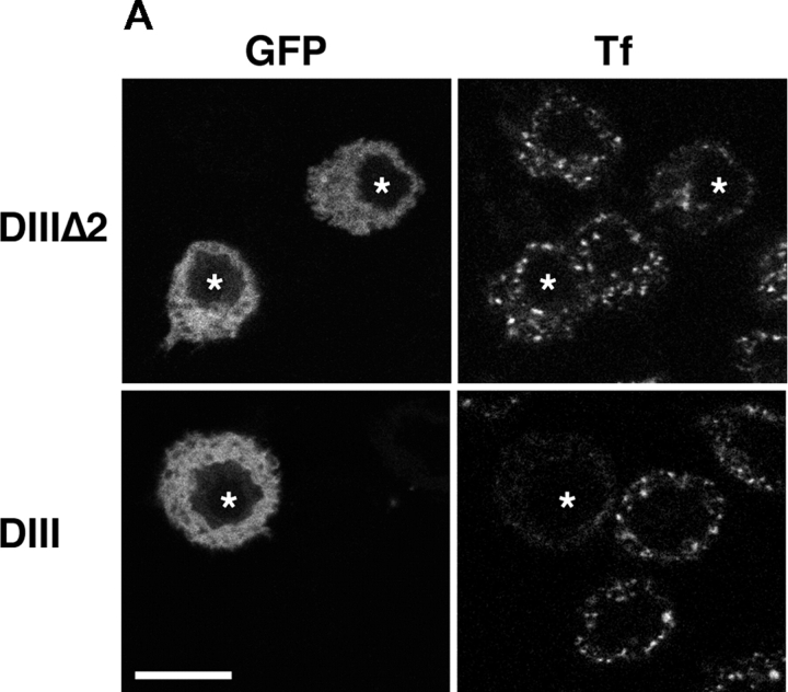
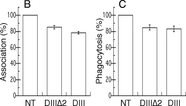
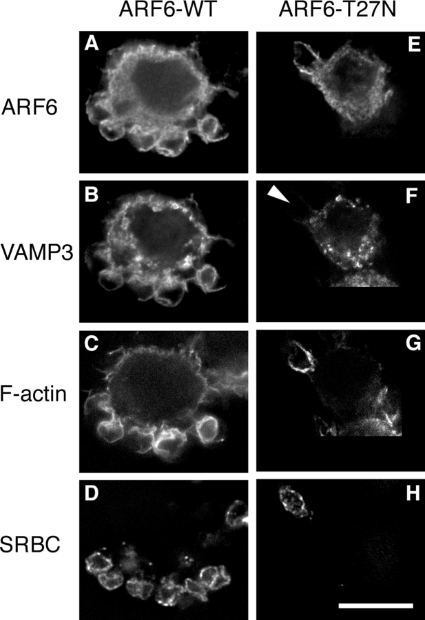
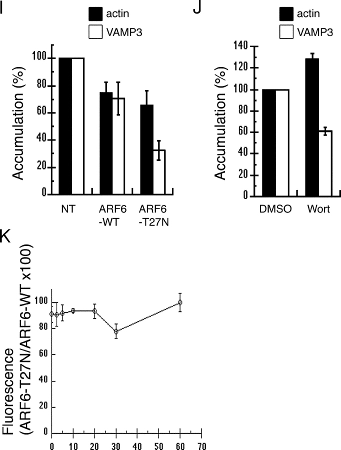
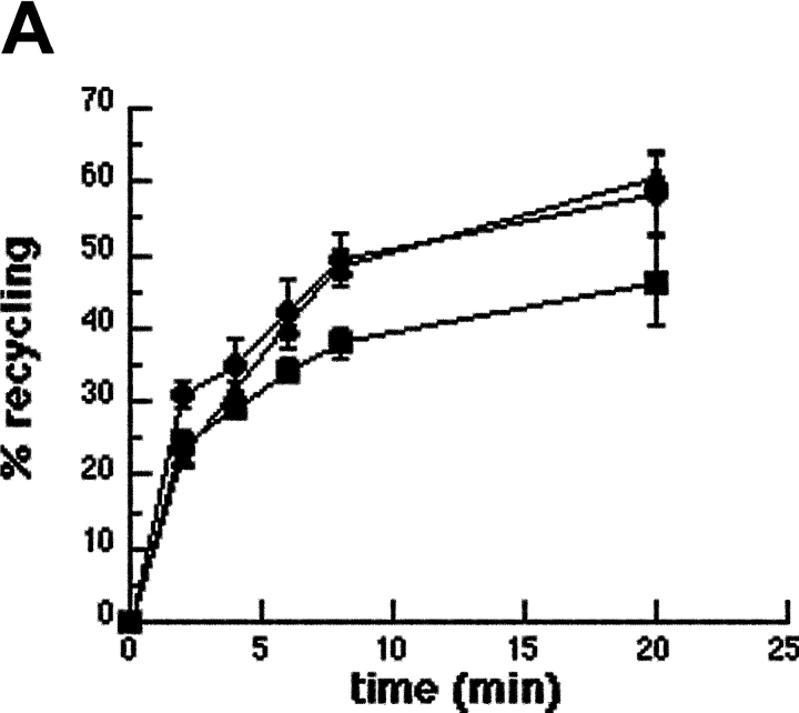
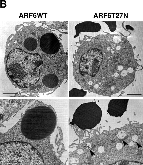
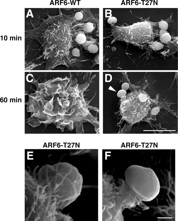
Engulfment of particles by phagocytes is induced by their interaction with specific receptors on the cell surface, which leads to actin polymerization and the extension of membrane protrusions to form a closed phagosome. Membrane delivery from internal pools is considered to play an important role in pseudopod extension during phagocytosis. Here, we report that endogenous ADP ribosylation factor 6 (ARF6), a small GTP-binding protein, undergoes a sharp and transient activation in macrophages when phagocytosis was initiated via receptors for the Fc portion of immunoglobulins (FcRs). A dominant-negative mutant of ARF6 (T27N mutation) dramatically affected FcR-mediated phagocytosis. Expression of ARF6-T27N lead to a reduction in the focal delivery of vesicle-associated membrane protein 3+ endosomal recycling membranes at phagocytosis sites, whereas actin polymerization was unimpaired. This resulted in an early blockade in pseudopod extension and accumulation of intracellular vesicles, as observed by electron microscopy. We conclude that ARF6 is a major regulator of membrane recycling during phagocytosis.


Figures

Figure 1.
Figure 1.
ARF6 is activated during phagocytosis. RAW264.7 macrophages transiently expressing ARF6-WT or ARF6-T27N were incubated with IgG-SRBC for 60 min at 37°C. The cells were fixed, and external SRBCs were stained with Cy3-coupled anti–rabbit IgG antibodies. After permeabilization, the expressed HA-tagged ARF6 constructs were detected by immunofluorescence. The efficiency of association (A) or phagocytosis (B) was calculated as indicated in Materials and methods. The mean ± SEM of three independent experiments is plotted. NT, non transfected. (C) ARF6 is transiently activated during phagocytosis. RAW264.7 macrophages were incubated with medium (control) for 10 min or with IgG-SRBC for various times at 37°C. Lysates were prepared and incubated with GST-GGA31–226 (top) or GST alone (middle). The bottom panel shows aliquots of total lysates. Western blot was performed with anti-ARF6 antibodies. Data are representative of three experiments. (D) Kinetics of activation of Rac and Cdc42 during phagocytosis. RAW264.7 macrophages were activated as described in C, and lysates were prepared and incubated with GST-CRIB. Western blot was performed with anti-Rac antibodies (top), then with anti-Cdc42 (bottom). Data are representative of four experiments.
Figure 2.
Figure 2.
An Eps15 mutant inhibits Tf endocytosis but not FcR-mediated phagocytosis. RAW264.7 cells transiently expressing GFP-Eps15DIIIΔ2 (top) or the dominant-negative construct GFP-Eps15DIII (bottom) were incubated for 30 min at 37°C in medium containing 60 nM Texas red-Tf. Cells were then fixed and analyzed by confocal microscopy. A medial optical section is shown. Left, GFP; right, Texas red-Tf. The asterisks indicate GFP-positive transfected cells. Bar, 10 μm. (B and C) Inhibition of receptor-mediated endocytosis does not block phagocytosis. Association (B) and phagocytic (C) efficiency of cells expressing GFP-Eps15DIIIΔ2 or GFP-Eps15DIII were compared with the efficiency of phagocytosis of nontransfected (NT) control cells, as described in Fig. 1 and Materials and methods. Data presented are the mean ± SEM of three independent experiments.
Figure 2.
Figure 2.
An Eps15 mutant inhibits Tf endocytosis but not FcR-mediated phagocytosis. RAW264.7 cells transiently expressing GFP-Eps15DIIIΔ2 (top) or the dominant-negative construct GFP-Eps15DIII (bottom) were incubated for 30 min at 37°C in medium containing 60 nM Texas red-Tf. Cells were then fixed and analyzed by confocal microscopy. A medial optical section is shown. Left, GFP; right, Texas red-Tf. The asterisks indicate GFP-positive transfected cells. Bar, 10 μm. (B and C) Inhibition of receptor-mediated endocytosis does not block phagocytosis. Association (B) and phagocytic (C) efficiency of cells expressing GFP-Eps15DIIIΔ2 or GFP-Eps15DIII were compared with the efficiency of phagocytosis of nontransfected (NT) control cells, as described in Fig. 1 and Materials and methods. Data presented are the mean ± SEM of three independent experiments.
Figure 3.
Figure 3.
ARF6-T27N inhibits the recruitment of VAMP3 (but not the polymerization of actin) at the site of phagocytosis. RAW264.7 cells stably expressing GFP-VAMP3 (B and F) were transiently transfected to express ARF6-WT-HA (A–D) or ARF6-T27N-HA (E–H) and were incubated with Cy5-IgG-SRBC (D and H) for 10 min at 37°C. The cells were then permeabilized and stained with Alexa® 350-phalloidin (C and G) to detect F-actin, and anti-HA followed by Cy3-labeled anti–rat IgG (A and E) to reveal HA-tagged ARF6. Cells were analyzed by conventional epifluorescence microscopy and deconvolution. One section is shown. Bar, 10 μm. Accumulations of GFP-VAMP3 (open bars) and polymerized actin (filled bars) were scored as described in Materials and methods for ARF6-WT– and ARF6-T27N–expressing cells (I) or for wortmannin-treated cells (J), and expressed as a percentage of the indexes obtained for control nontransfected (NT) cells (I) or DMSO-treated cells (J). Data are the mean ± SEM of at least four independent experiments performed with two different stable clones. Flow cytometry quantification of polymerized actin during phagocytosis (K). RAW264.7 cells positive for GFP, coexpressed with ARF6-WT or ARF6-T27N, were incubated with IgG-SRBC at 37°C and, at different time points, fixed, permeabilized, and stained with Alexa® 633–phalloidin. The levels of fluorescence were then measured by FACS® on at least 5,000 positive cells. The F-actin content of ARF6-T27N– expressing cells was then expressed as a percentage of the values obtained for ARF6-WT–expressing cells. Data are the mean ± SEM of four independent experiments performed.
Figure 3.
Figure 3.
ARF6-T27N inhibits the recruitment of VAMP3 (but not the polymerization of actin) at the site of phagocytosis. RAW264.7 cells stably expressing GFP-VAMP3 (B and F) were transiently transfected to express ARF6-WT-HA (A–D) or ARF6-T27N-HA (E–H) and were incubated with Cy5-IgG-SRBC (D and H) for 10 min at 37°C. The cells were then permeabilized and stained with Alexa® 350-phalloidin (C and G) to detect F-actin, and anti-HA followed by Cy3-labeled anti–rat IgG (A and E) to reveal HA-tagged ARF6. Cells were analyzed by conventional epifluorescence microscopy and deconvolution. One section is shown. Bar, 10 μm. Accumulations of GFP-VAMP3 (open bars) and polymerized actin (filled bars) were scored as described in Materials and methods for ARF6-WT– and ARF6-T27N–expressing cells (I) or for wortmannin-treated cells (J), and expressed as a percentage of the indexes obtained for control nontransfected (NT) cells (I) or DMSO-treated cells (J). Data are the mean ± SEM of at least four independent experiments performed with two different stable clones. Flow cytometry quantification of polymerized actin during phagocytosis (K). RAW264.7 cells positive for GFP, coexpressed with ARF6-WT or ARF6-T27N, were incubated with IgG-SRBC at 37°C and, at different time points, fixed, permeabilized, and stained with Alexa® 633–phalloidin. The levels of fluorescence were then measured by FACS® on at least 5,000 positive cells. The F-actin content of ARF6-T27N– expressing cells was then expressed as a percentage of the values obtained for ARF6-WT–expressing cells. Data are the mean ± SEM of four independent experiments performed.
Figure 4.
Figure 4.
Dominant-negative ARF6 inhibits membrane recycling. Macrophages transiently coexpressing ARF6-T27N and GFP or GFP alone were scraped, pelleted, and resuspended in the presence of Alexa® 633-Tf for 45 min at 37°C. The efflux of Tf was then followed by flow cytometry as described in Materials and methods. Cells positive for GFP coexpressed with ARF6-T27N (closed squares), cells expressing GFP alone (closed circles), as well as cells negative for GFP (closed triangles) were gated and analyzed. The plot represents the mean ± SEM of three independent experiments. (B) Macrophages expressing ARF6-T27N accumulate large intracellular vesicles. RAW264.7 macrophages transiently coexpressing ARF6-WT (left) or ARF6-T27N (right) with GFP were sorted by flow cytometry and incubated with IgG-SRBC for 20 min at 37°C, then fixed and processed for transmission EM. SRBCs appear as large electron-dense areas. Cells expressing ARF6-T27N exhibited enlarged electronlucent intracellular compartments, as assessed by direct counting of the number of characteristic enlarged compartments in 30 macrophages. The large intracellular compartments present in ARF6-T27N–positive cells often contained internal membranes (bottom right panel, arrowheads). Data are representative of two independent experiments (sorting and analysis). Bars, 2 μm.
Figure 4.
Figure 4.
Dominant-negative ARF6 inhibits membrane recycling. Macrophages transiently coexpressing ARF6-T27N and GFP or GFP alone were scraped, pelleted, and resuspended in the presence of Alexa® 633-Tf for 45 min at 37°C. The efflux of Tf was then followed by flow cytometry as described in Materials and methods. Cells positive for GFP coexpressed with ARF6-T27N (closed squares), cells expressing GFP alone (closed circles), as well as cells negative for GFP (closed triangles) were gated and analyzed. The plot represents the mean ± SEM of three independent experiments. (B) Macrophages expressing ARF6-T27N accumulate large intracellular vesicles. RAW264.7 macrophages transiently coexpressing ARF6-WT (left) or ARF6-T27N (right) with GFP were sorted by flow cytometry and incubated with IgG-SRBC for 20 min at 37°C, then fixed and processed for transmission EM. SRBCs appear as large electron-dense areas. Cells expressing ARF6-T27N exhibited enlarged electronlucent intracellular compartments, as assessed by direct counting of the number of characteristic enlarged compartments in 30 macrophages. The large intracellular compartments present in ARF6-T27N–positive cells often contained internal membranes (bottom right panel, arrowheads). Data are representative of two independent experiments (sorting and analysis). Bars, 2 μm.
Figure 5.
Figure 5.
ARF6-T27N inhibits membrane extension around the particles. RAW264.7 cells expressing ARF6-WT (A and C) or ARF6-T27N (B and D) were incubated with IgG-SRBC for 10 min (A and B) or 60 min (C and D) at 37°C, then fixed and processed for scanning EM. Arrowhead points to an abortive phagosome. Bar, 10 μm. (E and F) Scanning electron micrographs of macrophages expressing ARF6-T27N incubated with IgG-SRBC for 60 min at 37°C. Bar, 1 μm.

References

    1. Aderem, A. 2002. How to eat something bigger than your head. Cell. 110:5–8. - PubMed
    1. Aderem, A., and D.M. Underhill. 1999. Mechanisms of phagocytosis in macrophages. Annu. Rev. Immunol. 17:593–623. - PubMed
    1. Allen, L.A., C. Yang, and J.E. Pessin. 2002. Rate and extent of phagocytosis in macrophages lacking VAMP3. J. Leukoc. Biol. 72:217–221. - PMC - PubMed
    1. Altschuler, Y., S.-H. Liu, L. Katz, K. Tang, S. Hardy, F. Brodsky, G. Apodaca, and K. Mostov. 1999. ADP-ribosylation factor 6 and endocytosis at the apical surface of Madin-Darby canine kidney cells. J. Cell Biol. 147:7–12. - PMC - PubMed
    1. Araki, N., M.T. Johnson, and J.A. Swanson. 1996. A role for phosphoinositide 3-kinase in the completion of macropinocytosis and phagocytosis by macrophages. J. Cell Biol. 135:1249–1260. - PMC - PubMed

Publication types

MeSH terms