

DnaK-facilitated ribosome assembly in Escherichia coli revisited
- PMID: 12810912
- PMCID: PMC1370445
- DOI: 10.1261/rna.5360203
DnaK-facilitated ribosome assembly in Escherichia coli revisited
Abstract
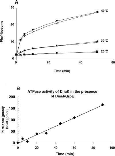
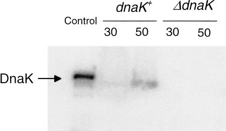
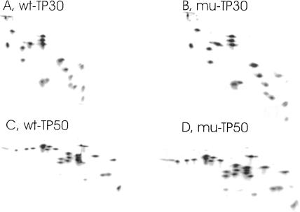
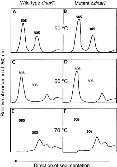
Assembly helpers exist for the formation of ribosomal subunits. Such a function has been suggested for the DnaK system of chaperones (DnaK, DnaJ, GrpE). Here we show that 50S and 30S ribosomal subunits from an Escherichia coli dnaK-null mutant (containing a disrupted dnaK gene) grown at 30 degrees C are physically and functionally identical to wild-type ribosomes. Furthermore, ribosomal components derived from mutant 30S and 50S subunits are fully competent for in vitro reconstitution of active ribosomal subunits. On the other hand, the DnaK chaperone system cannot circumvent the necessary heat-dependent activation step for the in vitro reconstitution of fully active 30S ribosomal subunits. It is therefore questionable whether the requirement for DnaK observed during in vivo ribosome assembly above 37 degrees C implicates a direct or indirect role for DnaK in this process.
Figures
Similar articles
-
Authentic precursors to ribosomal subunits accumulate in Escherichia coli in the absence of functional DnaK chaperone.Mol Microbiol. 2004 Jan;51(1):189-201. doi: 10.1046/j.1365-2958.2003.03813.x. Mol Microbiol. 2004. PMID: 14651621
-
The DnaK chaperone system facilitates 30S ribosomal subunit assembly.Mol Cell. 2002 Jul;10(1):129-38. doi: 10.1016/s1097-2765(02)00562-2. Mol Cell. 2002. PMID: 12150913
-
The chaperonin GroEL and other heat-shock proteins, besides DnaK, participate in ribosome biogenesis in Escherichia coli.Mol Gen Genet. 2001 Feb;264(6):796-808. doi: 10.1007/s004380000369. Mol Gen Genet. 2001. PMID: 11254127
-
Ribosome biogenesis is temperature-dependent and delayed in Escherichia coli lacking the chaperones DnaK or DnaJ.Mol Microbiol. 2009 Feb;71(3):748-62. doi: 10.1111/j.1365-2958.2008.06561.x. Epub 2008 Dec 11. Mol Microbiol. 2009. PMID: 19054328
-
The Escherichia coli chaperones involved in DNA replication.Philos Trans R Soc Lond B Biol Sci. 1993 Mar 29;339(1289):271-7; discussion 277-8. doi: 10.1098/rstb.1993.0025. Philos Trans R Soc Lond B Biol Sci. 1993. PMID: 8098531 Review.
Cited by
-
Nonbridging phosphate oxygens in 16S rRNA important for 30S subunit assembly and association with the 50S ribosomal subunit.RNA. 2005 May;11(5):657-67. doi: 10.1261/rna.7224305. Epub 2005 Apr 5. RNA. 2005. PMID: 15811917 Free PMC article.
-
Structural aspects of RbfA action during small ribosomal subunit assembly.Mol Cell. 2007 Nov 9;28(3):434-45. doi: 10.1016/j.molcel.2007.08.026. Mol Cell. 2007. PMID: 17996707 Free PMC article.
-
Ribosome biogenesis and the translation process in Escherichia coli.Microbiol Mol Biol Rev. 2007 Sep;71(3):477-94. doi: 10.1128/MMBR.00013-07. Microbiol Mol Biol Rev. 2007. PMID: 17804668 Free PMC article. Review.
-
Mutations of ribosomal protein S5 suppress a defect in late-30S ribosomal subunit biogenesis caused by lack of the RbfA biogenesis factor.RNA. 2015 Aug;21(8):1454-68. doi: 10.1261/rna.051383.115. Epub 2015 Jun 18. RNA. 2015. PMID: 26089326 Free PMC article.
-
Demonstration of the role of the DnaK chaperone system in assembly of 30S ribosomal subunits using a purified in vitro system.RNA. 2003 Dec;9(12):1418-21. doi: 10.1261/rna.5139703. RNA. 2003. PMID: 14623997 Free PMC article.
References
-
- Alix, J.-H. 1993. Extrinsic factors in ribosome assembly. In The translational apparatus: Structure, function, regulation, evolution (eds. K.H. Nierhaus et al.), pp. 173–184. Plenum, New York.
-
- Benaroudj, N., Fang, B., Triniolles, F., Ghelis, C., and Ladjimi, M.M. 1994. Overexpression in Escherichia coli, purification and characterization of the molecular chaperone HSC70. Eur. J. Biochem. 221: 121–128. - PubMed
-
- Blaha, G., Wilson, D.N., Stoller, G., Fischer, G., Willumeit, R., and Nierhaus, K.H. 2003. Localization of the trigger factor binding site on the ribosomal 50S subunit. J. Mol. Biol. 326: 887–897. - PubMed
-
- Bommer, U., Burkhardt, N., Jünemann, R., Spahn, C.M.T., Triana-Alonso, F.J., and Nierhaus, K.H. 1996. Ribosomes and polysomes. In Subcellular fractionation. A practical approach (eds. J. Graham and D. Rickwoods), pp. 271–301. IRL Press, Oxford University Press, Oxford, UK.
Publication types
MeSH terms
Substances
LinkOut - more resources
Full Text Sources
Other Literature Sources