. 2003 Jun 24;100(13):7988-95.
doi: 10.1073/pnas.1332808100. Epub 2003 Jun 16.

Mechanotransduction and flow across the endothelial glycocalyx

Affiliations

Mechanotransduction and flow across the endothelial glycocalyx

Sheldon Weinbaum et al. Proc Natl Acad Sci U S A. .

Abstract

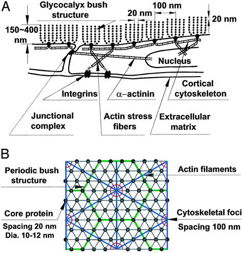
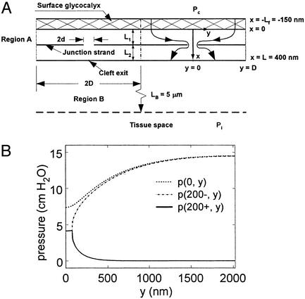
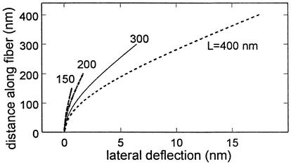
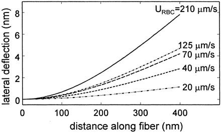
In this inaugural paper, we shall provide an overview of the endothelial surface layer or glycocalyx in several roles: as a transport barrier, as a porous hydrodynamic interface in the motion of red and white cells in microvessels, and as a mechanotransducer of fluid shearing stresses to the actin cortical cytoskeleton of the endothelial cell. These functions will be examined from a new perspective, the quasiperiodic ultrastructural model proposed in Squire et al. [Squire, J. M., Chew, M., Nneji, G., Neal, C., Barry, J. & Michel, C. (2001) J. Struct. Biol. 136, 239-255] for the 3D organization of the endothelial surface layer and its linkage to the submembranous scaffold. We shall show that the core proteins in the bush-like structures comprising the matrix have a flexural rigidity, EI, that is sufficiently stiff to serve as a molecular filter for plasma proteins and as an exquisitely designed transducer of fluid shearing stresses. However, EI is inadequate to prevent the buckling of these protein structures during the intermittent motion of red cells or the penetration of white cell microvilli. In these cellular interactions, the viscous draining resistance of the matrix is essential for preventing adhesive molecular interactions between proteins in the endothelial membrane and circulating cellular components.


Figures

Fig. 1.
Fig. 1.
(A) Sketch of ESL (not to scale) showing core protein arrangement and spacing of scattering centers along core proteins and their relationship to actin CC as proposed in ref. . (B) En face view of idealized model for core protein clusters and cluster foci and their relationship to hexagonal actin lattice in CC.
Fig. 2.
Fig. 2.
(A) Model geometry taken from ref. . (B) Pressure distribution behind ESL and fore and aft of TJ strand in mathematical model for flow through cleft of frog mesentery capillary.
Fig. 3.
Fig. 3.
Model predictions for lateral deflection of core proteins of different lengths L for a fluid shear stress of 10 dyn/cm2 at ESL edge. EI = 700 pN·nm2.
Fig. 4.
Fig. 4.
Model predictions for lateral deflection of core proteins beneath red cells moving at different velocities URBC. Spacing δ between ESL edge and red cell taken from measurements in ref. .
Fig. 5.
Fig. 5.
(A) Predictions of Eq. 12 for lateral tip deflection due to buckling of core proteins subject to a normal load P applied at their ends. δo is the initial unloaded tip displacement from vertical shown in Fig. 1B. Curve for δo = 0 is “elastica” theory prediction for large deflections (38). (B) Results in A converted to normal displacement of ESL. Right ordinate is the compressive force for 27-fiber model for core protein cluster in Fig. 1B.
Fig. 6.
Fig. 6.
Model prediction for time-dependent drainage of fluid from ESL after red cell arrest in a 5-μm-diameter capillary. A cell pressure of 2,420 dyn/cm2 is required to fully collapse the layer within 0.5 s at constant KP. This time is extended to 25 s if the variation of KP with compression is considered.

References

    1. Luft, J. H. (1966) Fed. Proc. 25, 1773-1783. - PubMed
    1. Vink, H. & Duling, B. R. (1996) Circ. Res. 79, 581-589. - PubMed
    1. Michel, C. C. (1997) Exp. Physiol. 82, 1-30. - PubMed
    1. Weinbaum, S. (1998) Ann. Biomed. Eng. 26, 627-643. - PubMed
    1. Hu, X. & Weinbaum, S. (1999) Microvasc. Res. 58, 281-304. - PubMed

Publication types

MeSH terms

LinkOut - more resources