

Case Reports
Diffusion-weighted imaging of cerebritis
Affiliations
- PMID: 12812934
- PMCID: PMC8149019
Item in Clipboard
Case Reports
Diffusion-weighted imaging of cerebritis
AJNR Am J Neuroradiol.
2003 Jun-Jul.
Abstract
Restricted water diffusion has been used to distinguish pyogenic abscess from other rim-enhancing brain masses; however diffusion-weighted imaging of cerebral infection before capsule formation has rarely been described. We report a case of fungal cerebritis in which water diffusion was more restricted than that of normal contralateral brain and the measured diffusion coefficient was in the range of that reported for pyogenic brain abscess. In the proper clinical setting, cerebritis should be considered in the differential diagnosis of an ill-defined focal brain mass associated with markedly restricted water diffusion.
Figures
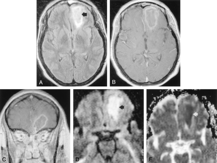
Images acquired on hospital day 5 during the early stage of left orbitofrontal cerebritis. A, FLAIR image (8000/105 [TR/TE]; TI, 2500) shows hyperintense subcortical white matter of left frontal lobe (arrow). B, Postcontrast T1-weighted image (650/17) shows no contrast enhancement around an ill-defined hypointense area (arrow). Note mucosal enhancement in left frontal sinus (white arrow). C, DW image (b = 1000 s/mm2) shows hyperintense signal (arrow) in frontal cerebritis. D, ADC map shows hypointense signal intensity (arrow) indicating restricted water diffusion. Mean ADC is 0.41 ± 0.04 (10−3mm2/s).
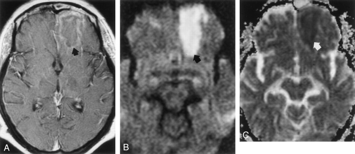
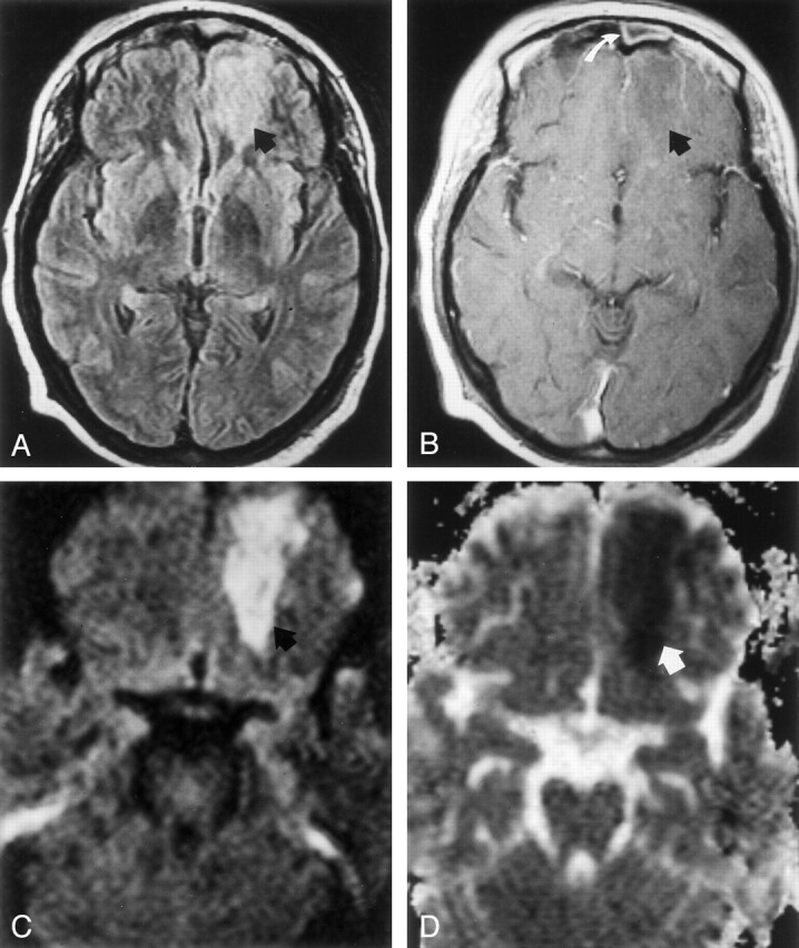
MR images acquired on hospital day 9 during the late stage of cerebritis or early stage of abscess. A, Contrast-enhanced T1-weighted image (650/17) shows thin, faint peripheral contrast enhancement around a left frontal mass (arrow). B, DW image (b = 1,000 s/mm2) demonstrates marked hyperintensity with a rim of even higher signal intensity (arrow). C, Restricted diffusion is indicated by hypointensity (arrow) on this ADC map. Mean ADC is 0.59 ± 0.03 (10−3mm2/s).
Images acquired before abscess aspiration on hospital day 15. A, FLAIR image (9000/105; TI; 2500) shows a poorly defined left frontal mass (arrow). B and C, Postcontrast axial (B) and coronal (C) T1-weighted images show a thin, well-defined enhancing wall, consistent with cerebral abscess. D, DW image (b = 1000 s/mm2) shows marked increased signal intensity is present in this mass (arrow). E, Marked hypointensity (arrow) on ADC map is consistent with restricted diffusion. Mean ADC is 0.56 ± 0.05 (10−3mm2/s).
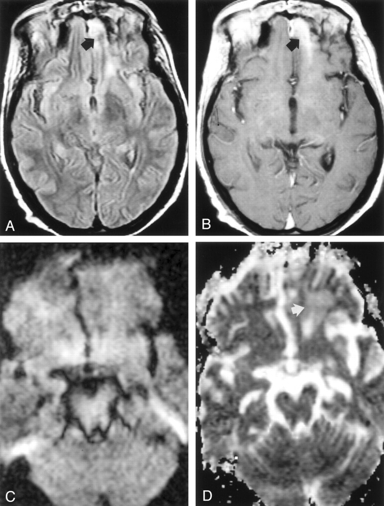
Images acquired after surgical aspiration and antifungal therapy; cerebral abscess has resolved, leaving focal gliosis. A, FLAIR (7000/105; TI, 2500) shows hyperintense signal in the left frontal lobe (arrow). B, Postcontrast T1-weighted image (650/17) shows a band of contrast enhancement at the site of treated abscess (arrow). C, DW image (b = 1000 s/mm2) shows symmetric signal intensities in frontal lobes. D, ADC map shows minimal hyperintensity (arrow) in left frontal lobe, a finding consistent with gliosis. Mean ADC is 1.87 ± 0.08 (10−3 m2/s).
References
-
- Schaefer P, Grant P, Gonzalez R. Diffusion-weighted MR imaging of the brain. Radiology 2000;217:331–345 - PubMed
-
- Ebisu T, Tanaka C, Umeda M, et al. Discrimination of brain abscess from necrotic or cystic tumors by diffusion-weighted echo planar imaging. Magn Reson Imaging 1996;14:1113–1116 - PubMed
-
- Noguchi K, Watanabe N, Nagayoshi T, et al. Role of diffusionweighted echo-planar MRI in distinguishing between brain abscess and tumour: a preliminary report. Neuroradiology 1999;41:171–174 - PubMed
-
- Tung G, Evangelista P, Rogg J, Duncan JI. Diffusion-weighted MR imaging of rim-enhancing brain masses: is markedly decreased water diffusion specific for brain abscess? AJR Am J Roentgenol 2001;177:709–712 - PubMed
Publication types
MeSH terms
LinkOut - more resources
Full Text Sources