. 2003 Jun-Jul;24(6):1123-9.

Total occlusion versus hairline residual lumen of the internal carotid arteries: accuracy of single section helical CT angiography

Affiliations

Total occlusion versus hairline residual lumen of the internal carotid arteries: accuracy of single section helical CT angiography

Michael H Lev et al. AJNR Am J Neuroradiol. 2003 Jun-Jul.

Abstract

Background and purpose: Routine carotid sonography and MR angiography cannot reliably detect the markedly reduced flow velocities associated with very severe carotid stenosis. In this study, we sought to evaluate the accuracy of single row detector helical CT angiography in distinguishing hairline residual lumen from total occlusion of severely stenosed internal carotid arteries (ICAs).

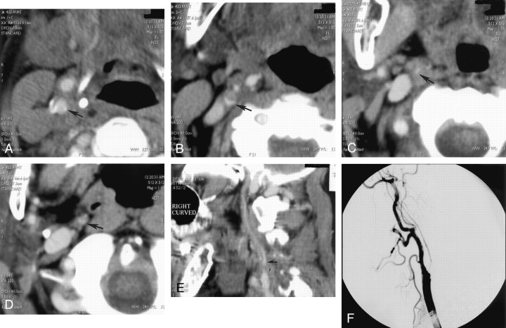
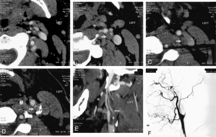
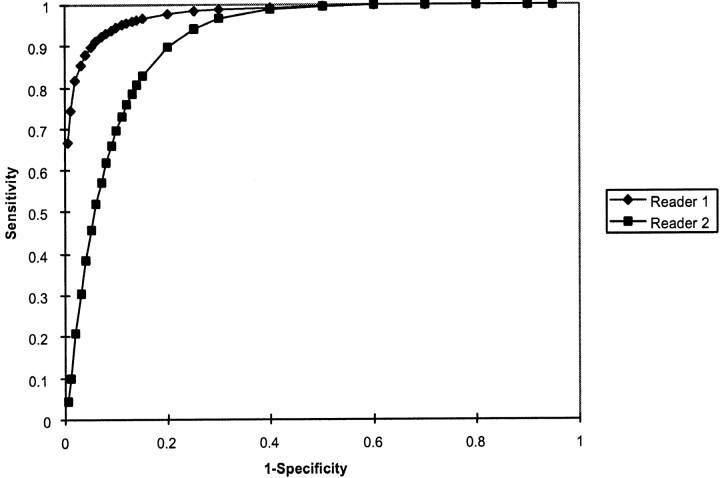
Methods: From our departmental data base of single row detector CT angiography studies performed for evaluation of ICA occlusive disease, 21 cases were identified with evidence of either hairline residual lumen or total occlusion on correlative conventional catheter radiographic arteriograms; these included seven cases of proved hairline residual lumen and 14 cases of proved total occlusion. Two neuroradiologists, blinded to the radiographic arteriography results, graded the diseased ICA on each CT angiogram as definitely occluded, probably occluded, indeterminate, probably patent, or definitely patent. Receiver operating characteristic curves were generated for each neuroradiologist.

Results: At an operating point on the receiver operating characteristic curve corresponding to 90% sensitivity, the first reader achieved 95% specificity and the second reader achieved 80% specificity for distinguishing hairline residual lumen from total occlusion. Absolute accuracy rates were 95% and 85%, respectively. No significant difference in accuracy was observed between the two readers (P =.28, two-tailed t test).

Conclusion: Single row detector CT angiography can distinguish total ICA occlusion from hairline residual lumen with a high degree of accuracy. In equivocal cases, conventional catheter arteriography may be desirable to confirm the diagnosis.


Figures

F<sc>ig</sc> 1.
Fig 1.
A−D, Selected axial view source scans from a CT angiogram of a 74-year-old female patient show continuity of the contrast-filled lumen (arrows) in consecutive axial views of the right ICA, reflecting a patent vascular lumen. Scans progress from inferior to superior levels, beginning at the patent carotid bifurcation. Note that in A, the jugular vein, posterior and lateral to the carotid artery, has not yet opacified. E, Sagittal view curved reformatted projection of this CT angiography dataset shows a hairline residual right ICA lumen with a slim sign extending into the petrous canal at the skull base. Note that the vessel appears falsely discontinuous along portions of the scan, which is a potential pitfall of this postprocessing technique, emphasizing the need for review of the axial view source scans. ICA origin is seen just superior to a discontinuous segment (arrow). F, Lateral view digital subtraction arteriogram obtained during injection of the right common carotid artery confirms critical stenosis of the right ICA origin, with diffuse narrowing (hairline residual lumen) of the more distal right ICA.
F<sc>ig</sc> 2.
Fig 2.
A−D, Selected source scans from a CT angiogram of a 45-year-old man. Unlike those shown in Figure 1, these scans reveal discontinuity of the intraluminal left ICA contrast column, reflecting an occluded vessel (arrow in A indicates residual ICA lumen; arrows in B−D indicate unopacified ICA lumen/enhancing vasa vasorum). Scans progress from inferior to superior levels. E, Curved reformatted projection constructed from this CT angiography dataset shows left ICA occlusion, with absent intraluminal contrast opacification. An opacified ascending pharyngeal artery (arrow) mimics a patent left ICA but cannot be followed into the petrous canal. F, Lateral view digital subtraction arteriogram obtained during injection of the left common carotid artery confirms total occlusion of the proximal left ICA.
F<sc>ig</sc> 3.
Fig 3.
Receiver operating characteristic curves for detection of hairline residual lumen by readers 1 and 2. Area under the receiver operating characteristic curve for reader 1 (which correlates with accuracy) was 0.98 ± 0.03; for reader 2, this area was 0.91 ± 0.06. No significant difference was observed between the accuracy of the two readers (P = .28, two-tailed t test). Of note, at an operating point corresponding to 90% sensitivity, reader 1 achieved 95% specificity and reader 2 achieved 80% specificity. At an operating point corresponding to a sensitivity of 95%, specificity was reduced to approximately 90% for reader 1 and 75% for reader 2.

Similar articles

Cited by

References

    1. Harrison MJ, Marshall J. Angiographic appearance of carotid bifurcation in patients with completed stroke, transient ischaemic attacks, and cerebral tumour. Br Med J 1976;1:205–207 - PMC - PubMed
    1. Thiele BL, Young JV, Chikos PM, Hirsch JH, Strandness DE Jr. Correlation of arteriographic findings and symptoms in cerebrovascular disease. Neurology 1980;30:1041–1046 - PubMed
    1. North American Symptomatic Carotid Endarterectomy Trialists’ Collaborative Group. The final results of the NASCET trial. N Engl J Med 1998;339:1415–1425 - PubMed
    1. Lippman HH, Sundt TM Jr., Holman CB. The poststenotic carotid slim sign: supurious internal carotid hypolasia. Mayo Clin Proc 1970;45:762–767 - PubMed
    1. O’Leary DH, Mattle H, Potter JE. Atheromatous pseudo-occlusion of the internal carotid artery. Stroke 1989;20:1168–1173 - PubMed

Publication types

LinkOut - more resources