

Case Reports
Sinonasal intestinal-type adenocarcinoma involvement of the paranasal sinuses
Affiliations
- PMID: 12812944
- PMCID: PMC8149027
Item in Clipboard
Case Reports
Sinonasal intestinal-type adenocarcinoma involvement of the paranasal sinuses
AJNR Am J Neuroradiol.
2003 Jun-Jul.
Abstract
We present a patient with a biopsy-proved sinonasal intestinal-type adenocarcinoma who presented with moderate confusion. He was found to have bifrontal hemorrhages, which to our knowledge has not been previously described in the literature for this entity. Intestinal-type adenocarcinoma should be in the differential diagnosis of aggressive lesions in the base of the skull with intracranial spread from the paranasal sinuses.
Figures
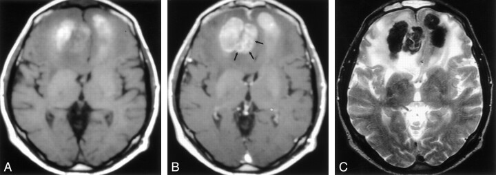
Axial MR images show a bifrontal lesion. A, T1-weighted (602/12/1 [TR/TE/NEX]) image shows the lesion with areas of high signal intensity. B, Postgadolinium T1-weighted (602/12/1) image shows enhancement of the paramidline portion of the lesion (arrows). C, T2-weighted (5000/96/1) image demonstrates areas of low signal intensity corresponding to the areas of high signal intensity on the T1-weighted images. Note the edema surrounding the lesion.

Sagittal MR images before (left) and after (right) the administration of a gadolinium-based contrast agent demonstrate an enhancing lesion extending from the ethmoid sinuses to the parenchyma of the frontal lobes.

Coronal postgadolinium T1-weighted (519/12/2) MR image shows the lesion extending from the ethmoid sinuses to the skull base and intracranially to the frontal lobes.
Axial CT scan demonstrates a bifrontal high-attenuating lesion.
References
-
- Weber AL, Strnton AC. Malignant tumors of the paranasal sinuses: radiologic, clinical, and histopathologic evaluation of 200 cases. Head Neck Surg 1984;6:761–776 - PubMed
-
- Lopez JI, Nevado M, Eizaguirre B, Perez A. Intestinal-type adenocarcinoma of the nasal cavity and paranasal sinuses: a clinicopathologic study of 6 cases. Tumori 1990;76:250–254 - PubMed
-
- Barnes L. Intestinal-type adenocarcinoma of the nasal cavity and paranasal sinuses. Am J Surg Pathol 1986;10:192–202 - PubMed
-
- Nylander LA, Demart JM. Carcinogenic effects of wood dust: review and discussion. Am J Ind Med 1993;24:619–647 - PubMed
Publication types
MeSH terms
LinkOut - more resources
Full Text Sources
Medical