

Treatment of wide-necked intracranial aneurysms with a self-expanding stent system: initial clinical experience
- PMID: 12812954
- PMCID: PMC8149018
Treatment of wide-necked intracranial aneurysms with a self-expanding stent system: initial clinical experience
Abstract
Background and purpose: Currently available stents for intracranial use usually are balloon-expandable coronary stents that carry the risk of damaging a dysplastic segment of the artery, with potential vessel rupture. We assessed the technical feasibility and efficacy of the combined application of a flexible, self-expanding neurovascular stent and detachable coils in the management of wide-necked intracranial aneurysms in humans.
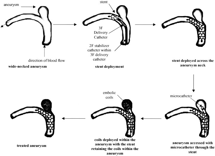
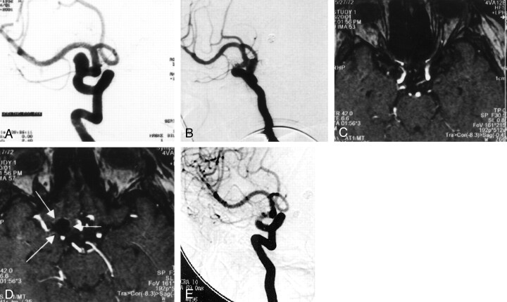
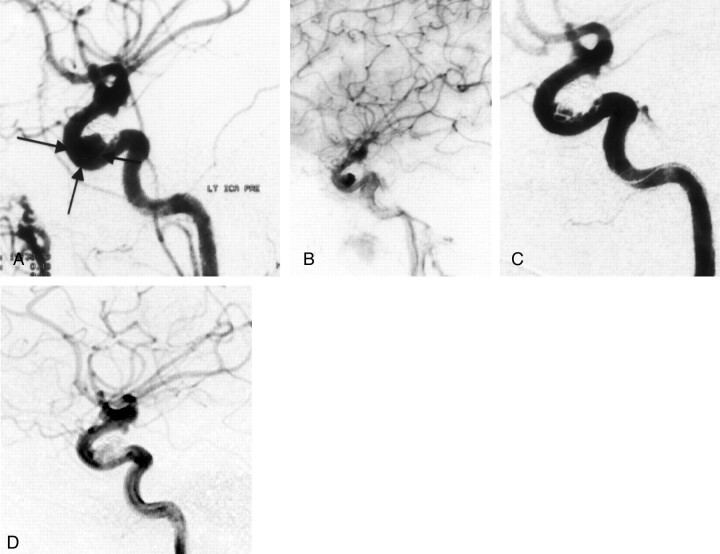
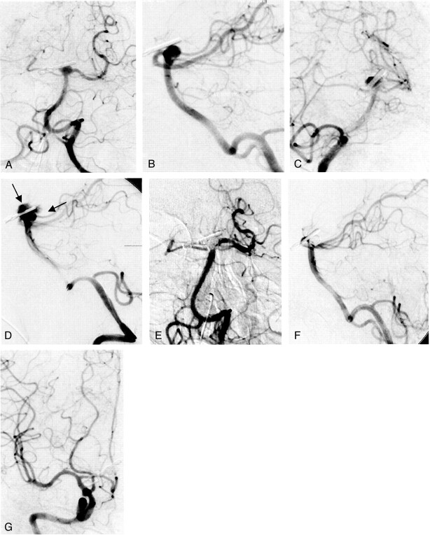
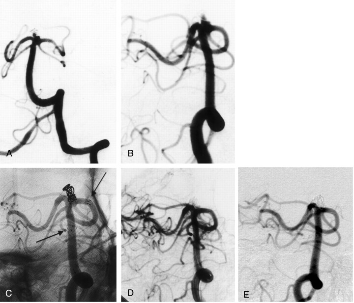
Methods: Four consecutive patients with a wide-necked intracranial aneurysm were treated with a combined approach that consisted of delivery of a flexible self-expanding neurovascular stent through a microcather to cover the neck of the aneurysm and subsequent filling of the aneurysm with coils through the stent interstices. The aneurysms were located at the internal carotid artery (n=2) and the basilar tip encroaching the P1 segment (n=2). Previous attempts with conventional endosaccular coil packing alone failed in all cases.
Results: Stent placement in the desired position with complete or nearly complete occlusion of the aneurysms was feasible in all patients. In one patient, aneurysm perforation with the microcatheter occurred and necessitated ventricular drainage, which led to a large parenchymal and intraventricular hemorrhage because of the strong anticoagulation regimen. Six-month follow-up demonstrated no focal neurologic sequelae in any of the patients, except slight memory dysfunction in the patient with bleeding.
Conclusion: Preliminary data demonstrate that this extremely flexible stent is technically easy to deploy and can be easily and safely maneuvered through severely tortuous vessels, enabling the treatment of intracranial wide-necked aneurysms. The combination of endovascular reconstruction of the parent vessel with use of a self-expanding stent followed by coil embolization offers a promising therapeutic alternative for wide-necked aneurysms not amenable to coil embolization alone. Although immediate angiographic results are promising, long-term angiographic and clinical follow-up is essential to determine permanent vessel patency and aneurysm occlusion rate.
Figures
Similar articles
-
Single-center experience with the Neuroform stent for endovascular treatment of wide-necked intracranial aneurysms.Surg Neurol. 2009 Dec;72(6):612-9. doi: 10.1016/j.surneu.2009.03.038. Epub 2009 Jul 14. Surg Neurol. 2009. PMID: 19604557
-
Immediate and midterm follow-up results of using an electrodetachable, fully retrievable SOLO stent system in the endovascular coil occlusion of wide-necked cerebral aneurysms.J Neurosurg. 2007 Jul;107(1):49-55. doi: 10.3171/JNS-07/07/0049. J Neurosurg. 2007. PMID: 17639873
-
Treatment of intracranial broad-neck aneurysms with a new self-expanding stent and coil embolization.AJNR Am J Neuroradiol. 2004 Apr;25(4):584-91. AJNR Am J Neuroradiol. 2004. PMID: 15090346 Free PMC article.
-
Intravascular stent and endovascular coil placement for a ruptured fusiform aneurysm of the basilar artery. Case report and review of the literature.J Neurosurg. 1997 Dec;87(6):944-9. doi: 10.3171/jns.1997.87.6.0944. J Neurosurg. 1997. PMID: 9384409 Review.
-
Comaneci Device for Temporary Coiling Assistance for Treatment of Wide-Necked Aneurysms: Initial Case Series and Systematic Literature Review.World Neurosurg. 2021 May;149:e85-e91. doi: 10.1016/j.wneu.2021.02.080. Epub 2021 Feb 25. World Neurosurg. 2021. PMID: 33640525
Cited by
-
Flat panel detector angiographic CT for stent-assisted coil embolization of broad-based cerebral aneurysms.AJNR Am J Neuroradiol. 2007 Nov-Dec;28(10):1902-8. doi: 10.3174/ajnr.A0697. Epub 2007 Sep 24. AJNR Am J Neuroradiol. 2007. PMID: 17893214 Free PMC article.
-
A review of technological innovations leading to modern endovascular brain aneurysm treatment.Front Neurol. 2023 Apr 11;14:1156887. doi: 10.3389/fneur.2023.1156887. eCollection 2023. Front Neurol. 2023. PMID: 37114225 Free PMC article. Review.
-
Iatrogenic carotid-cavernous fistula after stent assisted coil embolization of posterior communicating artery aneurysm.J Cerebrovasc Endovasc Neurosurg. 2015 Mar;17(1):43-8. doi: 10.7461/jcen.2015.17.1.43. Epub 2015 Mar 31. J Cerebrovasc Endovasc Neurosurg. 2015. PMID: 25874185 Free PMC article.
-
A novel self-expanding fully retrievable intracranial stent (SOLO): experience in nine procedures of stent-assisted aneurysm coil occlusion.Neuroradiology. 2006 Jul;48(7):471-8. doi: 10.1007/s00234-006-0062-7. Epub 2006 Jun 7. Neuroradiology. 2006. PMID: 16758153 Clinical Trial.
-
A novel flexible, retrievable endovascular stent system for small-vessel anatomy: preliminary in vivo data.AJNR Am J Neuroradiol. 2005 Apr;26(4):862-8. AJNR Am J Neuroradiol. 2005. PMID: 15814935 Free PMC article.
References
-
- Cognard C, Weill A, Spelle L, Piotin M, Castaings L, Rey A, Moret J. Long-term angiographic follow-up of 169 intracranial berry aneurysms occluded with detachable coils. Radiology 1999;212:348–356 - PubMed
-
- Byrne JV, Sohn MJ, Molyneux AJ, Chir B. Five-year experience in using coil embolization for ruptured intracranial aneurysms: outcomes and incidence of late rebleeding. J Neurosurg 1999;90:656–663 - PubMed
-
- Byrne JV, Bashiri M, Pasco A, Morris JH. A novel flexible endovascular stent for use in small and tortuous vessels. Neuroradiology 2000;42:56–61 - PubMed
Publication types
MeSH terms
LinkOut - more resources
Full Text Sources
Other Literature Sources
Medical