. 2003 Jun;111(12):1823-33.
doi: 10.1172/JCI16303.

Utilization of sialic acid as a coreceptor is required for reovirus-induced biliary disease

Affiliations

Utilization of sialic acid as a coreceptor is required for reovirus-induced biliary disease

Erik S Barton et al. J Clin Invest. 2003 Jun.

Abstract

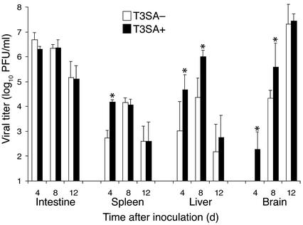
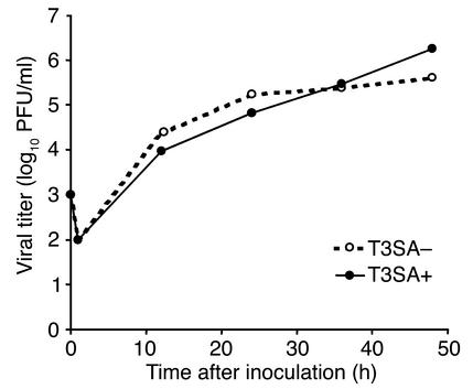
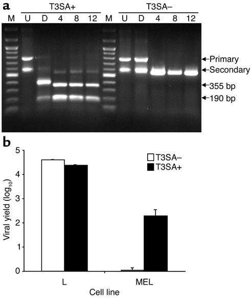
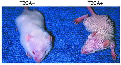
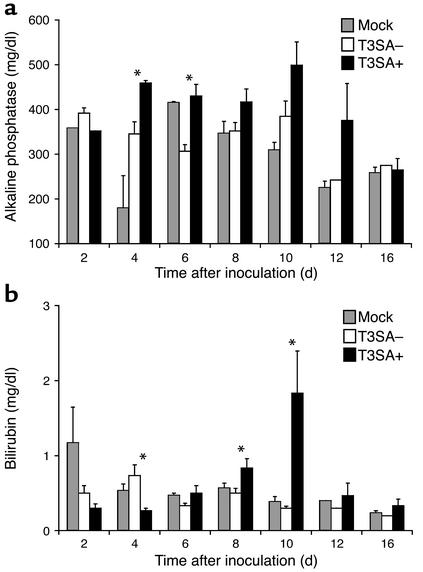
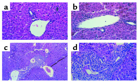
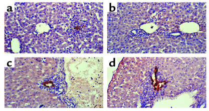
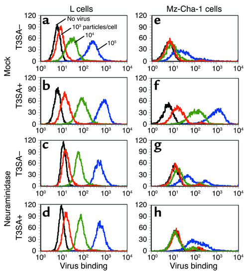
Infection of neonatal mice with some reovirus strains produces a disease similar to infantile biliary atresia, but previous attempts to correlate reovirus infection with this disease have yielded conflicting results. We used isogenic reovirus strains T3SA- and T3SA+, which differ solely in the capacity to bind sialic acid as a coreceptor, to define the role of sialic acid in reovirus encephalitis and biliary tract infection in mice. Growth in the intestine was equivalent for both strains following peroral inoculation. However, T3SA+ spread more rapidly from the intestine to distant sites and replicated to higher titers in spleen, liver, and brain. Strikingly, mice infected with T3SA+ but not T3SA- developed steatorrhea and bilirubinemia. Liver tissue from mice infected with T3SA+ demonstrated intense inflammation focused at intrahepatic bile ducts, pathology analogous to that found in biliary atresia in humans, and high levels of T3SA+ antigen in bile duct epithelial cells. T3SA+ bound 100-fold more efficiently than T3SA- to human cholangiocarcinoma cells. These observations suggest that the carbohydrate-binding specificity of a virus can dramatically alter disease in the host and highlight the need for epidemiologic studies focusing on infection by sialic acid-binding reovirus strains as a possible contributor to the pathogenesis of neonatal biliary atresia.


Figures

Figure 1
Figure 1
Growth of T3SA– and T3SA+ in mice after peroral inoculation. ND4 Swiss Webster mice, 2–3 days old, were inoculated perorally with 2.5 × 10³ PFU of either T3SA– or T3SA+ in a volume of 50 μl. At the indicated times after inoculation, mice were euthanized and organs were collected. Organs were homogenized by sonication, and titers of virus present in homogenates were determined by plaque assay. Each bar represents the average viral titer from three mice. Error bars indicate SEM. *P < 0.05 by Student t test.
Figure 2
Figure 2
Growth of T3SA– and T3SA+ in mice after intracranial inoculation. ND4 Swiss Webster mice, 2–3 days old, were inoculated intracranially with 1 × 10³ PFU of either T3SA– or T3SA+ in a volume of 5 μl. At the indicated times after inoculation, mice were euthanized and brains were collected. Brain tissue was homogenized by sonication, and titers of virus present in homogenates were determined by plaque assay. Each data point represents the average viral titer from two to four brains.
Figure 3
Figure 3
Genotypic and phenotypic characterization of virus isolated from mice following infection with T3SA– and T3SA+. (a) Viral genomic RNA was extracted from either homogenized livers of infected mice or second-passage lysate stocks. S1 gene segment cDNA’s were amplified using RT-PCR (labeled at right as Primary and Secondary), and restriction enzyme digestion was performed using BstNI. Digested secondary PCR products (labeled at right as 355 bp and 190 bp) were visualized by agarose gel electrophoresis and ethidium bromide staining. Undigested (U) and digested (D) stock virions are shown along with infected livers from days 4, 8, and 12 after inoculation. The lanes labeled M were loaded with 100-bp markers. (b) Homogenized livers obtained 4, 8, and 12 days after inoculation were passaged in L cells for 72 hours. L cell lysate (150 μl) was adsorbed to either L cells or MEL cells (2 × 10⁵) and incubated at 37°C for 24 hours. Viral titers at 0 hours and 24 hours were determined from liver samples obtained from three to four mice by plaque assay using L cells. Viral yields were calculated by dividing titer at 24 hours by titer at 0 hours, and average viral yields for all at 3 days after inoculation are shown. Error bars indicate SEM.
Figure 4
Figure 4
Mice infected with T3SA+ develop OHE. Two-day-old ND4 Swiss Webster mice were inoculated perorally with 1 × 10⁴ PFU of either T3SA– or T3SA+ and photographed 11 days after inoculation.
Figure 5
Figure 5
Serum alkaline phosphatase and bilirubin levels in mice after infection with T3SA– and T3SA+. ND4 Swiss Webster mice, 2–3 days old, were inoculated perorally with 2.5 × 10³ PFU of either T3SA– or T3SA+. Mock-infected animals were inoculated perorally with PBS. At the indicated times after inoculation, mice were euthanized, blood was collected, and serum was separated by centrifugation. Total alkaline phosphatase (a) and bilirubin (b) levels were determined for each serum sample. Each bar represents an average enzyme level for one to four mice. Error bars indicate SEM. *P < 0.05, T3SA– vs. T3SA+ by Student t test.
Figure 6
Figure 6
Liver histopathology in mice following infection with T3SA– and T3SA+. ND4 Swiss Webster mice, 2–3 days old, were inoculated perorally with PBS (a) or 2.5 × 10³ PFU of either T3SA– (b) or T3SA+ (c and d). At 8 days after inoculation, liver tissue was harvested, embedded in paraffin, thin-sectioned, and stained with hematoxylin and eosin. Sections from approximately ten mice infected with each virus were examined and showed qualitatively similar results. Photomicrographs were prepared at final magnifications of ×100 (c) or ×400 (a, b, and d).
Figure 7
Figure 7
Immunohistochemical localization of reovirus antigen in bile duct epithelial cells. ND4 Swiss Webster mice, 2–3 days old, were inoculated perorally with 2.5 × 10³ PFU of either T3SA– (a and b) or T3SA+ (c and d). Six days after inoculation, liver tissue was harvested, embedded in paraffin, thin-sectioned, and stained for reovirus antigen using rabbit anti-reovirus serum and horseradish peroxidase. Dark brown staining indicates reovirus antigen. Sections from approximately ten mice infected with each virus were examined and showed qualitatively similar results. Photomicrographs were prepared at ×400 final magnification. Representative sections from two separate animals are shown.
Figure 8
Figure 8
Binding of T3SA– and T3SA+ to L cells and Mz-Cha-1 cells. L cells (ad) or Mz-Cha-1 biliary epithelial cells (eh) were either mock-treated (a, b, e, and f) or treated with C. perfringens neuraminidase to remove cell-surface terminal sialic acid residues (c, d, g, and h). Cells were incubated with 0 (black line), 103 (red line), 104 (green line), or 105 (blue line) particles per cell of T3SA– (a, c, e, and g) or T3SA+ (b, d, f, and h). Unbound virus was removed, and bound virus on the cell surface was detected using rabbit anti-reovirus serum followed by phycoerythrin-labeled goat anti-rabbit Fab’s. Histograms represent data gated on total live-cell populations. Shown are representative histograms from one experiment (L cells) or two experiments (Mz-Cha-1 cells). Preimmune rabbit serum caused less than a threefold increase in fluorescence in the presence of 106 particles per cell of virus (data not shown).

References

    1. Tyler, K.L. 2001. Mammalian reoviruses. In Fields virology. 4th edition. D.M. Knipe and P.M. Howley, editors. Lippincott-Raven Publishers. Philadelphia, Pennsylvania, USA. 1729–1945.
    1. Virgin, H.W., Tyler, K.L., and Dermody, T.S. 1997. Reovirus. In Viral pathogenesis. N. Nathanson, editor. Lippincott-Raven Publishers. Philadelphia, Pennsylvania, USA. 669–699.
    1. Tardieu M, Weiner HL. Viral receptors on isolated murine and human ependymal cells. Science. 1982;215:419–421. - PubMed
    1. Morrison LA, Sidman RL, Fields BN. Direct spread of reovirus from the intestinal lumen to the central nervous system through vagal autonomic nerve fibers. Proc. Natl. Acad. Sci. U. S. A. 1991;88:3852–3856. - PMC - PubMed
    1. Kaye KM, Spriggs DR, Bassel-Duby R, Fields BN, Tyler KL. Genetic basis for altered pathogenesis of an immune-selected antigenic variant of reovirus type 3 (Dearing) J. Virol. 1986;59:90–97. - PMC - PubMed

Publication types

MeSH terms