. 2003 Jun;111(12):1835-42.
doi: 10.1172/JCI17455.

Knockout of insulin and IGF-1 receptors on vascular endothelial cells protects against retinal neovascularization

Affiliations

Knockout of insulin and IGF-1 receptors on vascular endothelial cells protects against retinal neovascularization

Tatsuya Kondo et al. J Clin Invest. 2003 Jun.

Abstract

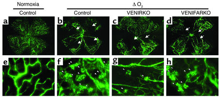
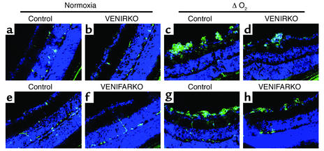
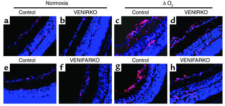
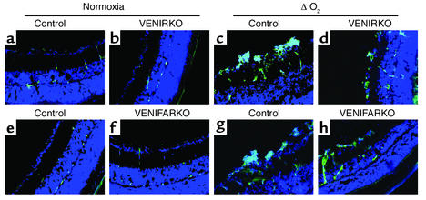
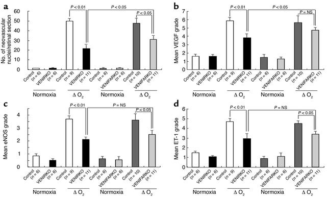
Both insulin and IGF-1 have been implicated in control of retinal endothelial cell growth, neovascularization, and diabetic retinopathy. To precisely define the role of insulin and IGF-1 signaling in endothelium in these processes, we have used the oxygen-induced retinopathy model to study mice with a vascular endothelial cell-specific knockout of the insulin receptor (VENIRKO) or IGF-1 receptor (VENIFARKO). Following relative hypoxia, VENIRKO mice show a 57% decrease in retinal neovascularization as compared with controls. This is associated with a blunted rise in VEGF, eNOS, and endothelin-1. By contrast, VENIFARKO mice show only a 34% reduction in neovascularization and a very modest reduction in mediator generation. These data indicate that both insulin and IGF-1 signaling in endothelium play a role in retinal neovascularization through the expression of vascular mediators, with the effect of insulin being most important in this process.


Figures

Figure 1
Figure 1
Retinal flat-mount pictures of VENIRKO and VENIFARKO mice exposed to hypoxia show decreased loss of blood vessels and fewer vascular tufts. Controls (b and f), VENIRKO (c and g), and VENIFARKO (d and h) mice were placed in a 75% oxygen environment from P7 to P12, then returned to normoxia for 5 days. Control in normoxia sample is shown in a and e. Retinal vessels were visualized by fluorescein-conjugated dextran injection at P17. Control in normoxia shows no central avascular area (a) and no peripheral tuft formation (e). Low magnification of retina from control mice exposed to hypoxia (b) shows large central avascular areas (indicated by arrows). High magnifications of these retinas (f) reveal neovascular tuft formation (arrowheads). Retinas of VENIRKO mice (c and g) show smaller avascular areas and a reduction of tufts. Retinas of VENIFARKO mice (d and h) show intermediate central avascular areas and tufts between control and VENIRKO mice. These flat-mount pictures are representative of at least six similar experiments.
Figure 2
Figure 2
Retinas of VENIRKO and VENIFARKO mice exposed to hypoxia show fewer neovascular nuclei on PAS and hematoxylin staining. Frozen sections (6 μm) of retinas were stained using PAS and hematoxylin. Retinas prepared from each control (a and e), VENIRKO (b), and VENIFARKO (f) mouse exposed in normoxia showed no neovascularization. Sections from each control mouse in the ΔO2 model (c and g) had significant numbers of protruding nuclei above the inner limiting membrane, whereas the number of nuclei in VENIRKO retinas after the same treatment (d) was decreased 57% as compared with controls (P < 0.01; see Figure 6a). Furthermore, the number of nuclei in VENIFARKO retinas (h) was reduced 34%, as in control mice (P < 0.05; see Figure 6a).
Figure 3
Figure 3
VEGF immunoreactivity in control retina is induced under hypoxia, but is less in VENIFARKO and even less in VENIRKO mice. VEGF immunoreactivity was constitutively observed in each control (a and e), VENIRKO (b), and VENIFARKO (f) retina and was highly induced in the ΔO2 model of each control in the preretinal area (c and g). The induction of VEGF in VENIRKO retina (d) was blunted 34% (P < 0.01; see Figure 6b), and that in VENIFARKO retina (h) was reduced 18% (not significant; see Figure 6b).
Figure 4
Figure 4
The eNOS immunoreactivity in control retina is increased under hypoxia, but is less in VENIFARKO and even less in VENIRKO mice. The eNOS protein was expressed in each control (a and e), VENIRKO (b), and VENIFARKO (f) retina and was strongly increased when exposed to relative hypoxic conditions in the microvascular preretinal area in each control (c and g). The induction of eNOS in VENIRKO retina (d) was decreased 43% (P < 0.01; see Figure 6c), and that in VENIFARKO retina (h) was reduced 31% (P < 0.05; see Figure 6c).
Figure 5
Figure 5
ET-1 immunoreactivity in control retina is upregulated under hypoxia, but is less in VENIFARKO and even less in VENIRKO mice. ET-1 immunoreactivity was constitutively seen in each control (a and e), VENIRKO (b), and VENIFARKO (f) retina and was obviously upregulated in the ΔO2 model of each control in the preretinal region (c and g). The induction of ET-1 in VENIRKO retina (d) was blunted 37% (P < 0.01; see Figure 6d), and that in VENIFARKO retina (h) declined 24% (P < 0.05; see Figure 6d).
Figure 6
Figure 6
Quantitative results for suppression of neovascularization and VEGF, eNOS, and ET-1 immunoreactivity. (a) The number of neovascular nuclei per section is shown. Quantitative (b) VEGF, (c) eNOS, and (d) ET-1 immunoreactive intensity are shown.

Comment in

  • An eye on insulin.
    Bronson SK, Reiter CE, Gardner TW. Bronson SK, et al. J Clin Invest. 2003 Jun;111(12):1817-9. doi: 10.1172/JCI18927. J Clin Invest. 2003. PMID: 12813016 Free PMC article. Review.

References

    1. Moss SE, Klein R, Klein BE. The 14-year incidence of visual loss in a diabetic population. Ophthalmology. 1998;105:998–1003. - PubMed
    1. Koya D, King GL. Protein kinase C activation and the development of diabetic complications. Diabetes. 1998;47:859–866. - PubMed
    1. Katzman GH. Retinopathy of prematurity: is suppression of neovascularization achievable? J. Pediatr. 1996;129:618–619. - PubMed
    1. Aiello LP, et al. Diabetic retinopathy. Diabetes Care. 1998;21:143–156. - PubMed
    1. Hamanaka T, Akabane N, Yajima T, Takahashi T, Tanabe A. Retinal ischemia and angle neovascularization in proliferative diabetic retinopathy. Am. J. Ophthalmol. 2001;132:648–658. - PubMed

Publication types

MeSH terms