. 2003 Jun;111(12):1923-31.
doi: 10.1172/JCI15722.

Altered cutaneous immune parameters in transgenic mice overexpressing viral IL-10 in the epidermis

Affiliations

Altered cutaneous immune parameters in transgenic mice overexpressing viral IL-10 in the epidermis

Wanhong Ding et al. J Clin Invest. 2003 Jun.

Abstract

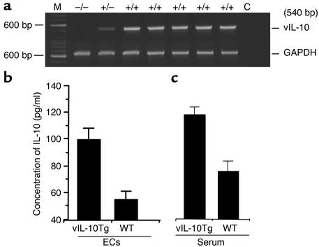
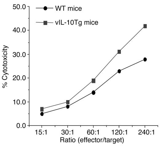
IL-10 is a pleiotropic cytokine that inhibits several immune parameters, including Th1 cell-mediated immune responses, antigen presentation, and antigen-specific T cell proliferation. Recent data implicate IL-10 as a mediator of suppression of cell-mediated immunity induced by exposure to UVB radiation (280-320 nm). To investigate the effects of IL-10 on the cutaneous immune system, we engineered transgenic mice that overexpress viral IL-10 (vIL-10) in the epidermis. vIL-10 transgenic mice demonstrated a reduced number of I-A(+) epidermal and dermal cells and fewer I-A(+) hapten-bearing cells in regional lymph nodes after hapten painting of the skin. Reduced CD80 and CD86 expression by I-A(+) epidermal cells was also observed. vIL-10 transgenic mice demonstrated a smaller delayed-type hypersensitivity response to allogeneic cells upon challenge but had normal contact hypersensitivity to an epicutaneously applied hapten. Fresh epidermal cells from vIL-10 transgenic mice showed a decreased ability to stimulate allogeneic T cell proliferation, as did splenocytes. Additionally, chronic exposure of mice to UVB radiation led to the development of fewer skin tumors in vIL-10 mice than in WT controls, and vIL-10 transgenic mice had increased splenic NK cell activity against YAC-1targets. These findings support the concept that IL-10 is an important regulator of cutaneous immune function.


Figures

Figure 1
Figure 1
ECs from vIL-10Tg mice express mRNA for vIL-10 and secrete vIL-10 protein. (a) RT-PCR. Lane 1, DNA standard; lane 2, WT mice; lane 3, heterozygote transgenic mouse; lanes 4–8, homozygote transgenic mice; lane 9, RNA/PCR without RT. (b) vIL-10 in supernatants from fresh EC cultures and serum was measured by ELISA. (c) vIL-10 could be detected in serum from vIL-10Tg mice. n = 5, both groups. Error bars represent ± SE. (b) P < 0.0005, vIL-10Tg vs. WT; (c) P = 0.0005, vIL-10Tg vs. WT.
Figure 2
Figure 2
I-A+ expression is reduced in the epidermis and dermis of vIL-10Tg, although a normal proportion of ATPase+ cells is present. (a) FACS analysis of I-A+ ECs from a single transgenic and a single WT mouse. (b) Comparison of mean percentage of I-Aq+ cells from groups of ten WT mice and ten vIL-10Tg mice. P = 0.0001. (c) Mean intensity of I-Aq expression of vIL-10Tg mice and WT mice. n = 5, both groups; P = 0.0124. (d) Frozen sections of truncal skin from vIL-10Tg and WT mice were prepared, and staining for I-Aq was carried out. Decreased numbers of I-Aq cells were seen in the dermis of vIL-10Tg mice. (e) Normal numbers of ATPase+ cells are present in vIL-10Tg mice. Epidermal sheets were prepared from the dorsum of vIL-10Tg mice and WT mice and stained for ATPase activity.
Figure 3
Figure 3
Skin painting with FITC yields fewer hapten-bearing I-A+ cells in draining lymph nodes. vIL-10Tg and WT mice were painted on the thorax and abdomen with FITC, and 18 hours later, inguinal and axillary lymph nodes were collected. DCs were isolated, and two-color analysis for I-Aq and FITC was performed. (a) The difference in frequency of FITC-bearing cells from a single vIL-10Tg mouse and a single WT mouse. (b) Comparison of mean frequency of hapten-bearing I-A+ cells from groups of seven vIL-10Tg mice and seven WT mice. Error bars represent ± SEM. P = 0.006, vIL-10Tg vs. WT.
Figure 4
Figure 4
Cultured ECs from vIL-10Tg mice have decreased expression of CD80 and CD86. Fresh ECs were prepared from both vIL-10Tg and WT mice, cultured for 72 hours in vitro, and then stained with PE-labeled anti-CD80 or anti-CD86. The mean fluorescence intensity of CD80 (a) and CD86 (b) was assessed. n = 5. (a) P = 0.0008, vIL-10Tg vs. WT. (b) P = 0.03, vIL-10Tg vs. WT.
Figure 5
Figure 5
ECs and splenocytes from vIL-10Tg mice have decreased ability to stimulate allogeneic T cell proliferation. (a) Various numbers of ECs (stimulators; S) were prepared from vIL-10Tg mice and WT mice. These cells were cocultured with 2 × 10⁵ nylon wool-purified allogeneic (BALB/c) T cells (responders; R). Control ECs and T cells were cultured alone. T cell proliferation was measured by 3H-thymidine uptake. Data are expressed as the mean cpm of triplicate cultures ± SEM. (b) Various numbers of splenocytes (stimulators; S) were prepared from vIL-10Tg mice and WT mice and x-irradiated. These cells were cocultured with 2 × 10⁵ allogeneic (BALB/c) splenocytes (responders; R). Stimulators and responders were cultured alone as controls. T cell proliferation was measured by 3H-thymidine uptake. Data are expressed as the mean cpm of triplicate cultures ± SEM.
Figure 6
Figure 6
A decreased DTH response was observed in vIL-10Tg mice. WT mice (groups A and C) and vIL-10Tg mice (groups B and D) were examined. Groups of vIL-10Tg and WT mice were injected subcutaneously with splenocytes from naive allogeneic CAF1 mice (groups C and D) or were not immunized (groups A and B, negative control groups). Six days after immunization, all mice were challenged by subcutaneous injection of the left hind footpad with CAF1 splenocytes and 24-hour footpad swelling was assessed as a measure of DTH response. Error bars represent ± SEM. P = 0.007, C vs. D; P = NS, A vs. B.
Figure 7
Figure 7
vIL-10Tg mice develop fewer skin tumors in response to chronic UVB irradiation. Groups of WT and vIL-10Tg mice were exposed to 2.16 kJ/m2 of UVB irradiation from FS40 sunlamps three times per week for 27 weeks. Significantly fewer tumors developed over time in vIL-10Tg mice than in WT control mice (P < 0.05; n = 20, both groups).
Figure 8
Figure 8
Splenocytes from vIL-10Tg mice demonstrate enhanced NK cell activity compared with splenocytes from WT mice. Varying numbers of splenocytes (effector cells) from vIL-10Tg and WT mice were incubated with YAC-1 cells (target cells) for 4 hours in round-bottomed 96-well plates. Cells from vIL-10Tg mice exhibited significantly greater NK cell activity than cells from WT mice (P = 0.01).

References

    1. Taga K, Tosato G. IL-10 inhibits human T cell proliferation and IL-2 production. J. Immunol. 1992;148:1143–1148. - PubMed
    1. Del Prete G, et al. Human IL-10 is produced by both type 1 helper (Th1) and type 2 helper (Th2) T cell clones and inhibits their antigen-specific proliferation and cytokine production. J. Immunol. 1993;150:353–360. - PubMed
    1. Li L, Elliott JF, Mosmann TR. IL-10 inhibits cytokine production, vascular leakage, and swelling during T helper 1 cell-induced delayed-type hypersensitivity. J. Immunol. 1994;153:3967–3978. - PubMed
    1. Macatonia SE, Doherty TM, Knight SC, O’Garra A. Differential effect of IL-10 on dendritic cell-induced T cell proliferation and IFN-gamma production. J. Immunol. 1993;150:3755–3765. - PubMed
    1. Enk AH, Angeloni VL, Udey MC, Katz SI. Inhibition of Langerhans cell antigen-presenting function by IL-10. A role for IL-10 in induction of tolerance. J. Immunol. 1993;151:2390–2398. - PubMed

Publication types

MeSH terms