. 2003 Jul;185(13):3804-12.
doi: 10.1128/JB.185.13.3804-3812.2003.

Molecular analysis of the copper-transporting efflux system CusCFBA of Escherichia coli

Affiliations

Molecular analysis of the copper-transporting efflux system CusCFBA of Escherichia coli

Sylvia Franke et al. J Bacteriol. 2003 Jul.

Abstract

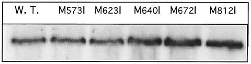
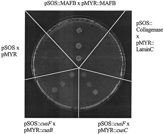
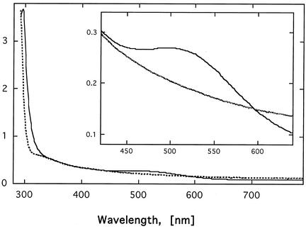
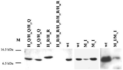
The cus determinant of Escherichia coli encodes the CusCFBA proteins that mediate resistance to copper and silver by cation efflux. CusA and CusB were essential for copper resistance, and CusC and CusF were required for full resistance. Replacements of methionine residues 573, 623, and 672 with isoleucine in CusA resulted in loss of copper resistance, demonstrating their functional importance. Substitutions for several other methionine residues of this protein did not have any effect. The small 10-kDa protein CusF (previously YlcC) was shown to be a periplasmic protein. CusF bound one copper per polypeptide. The pink CusF copper protein complex exhibited an absorption maximum at around 510 nm. Methionine residues of CusF were involved in copper binding as shown by site-directed mutagenesis. CusF interacted with CusB and CusC polypeptides in a yeast two-hybrid assay. In contrast to other well-studied CBA-type heavy metal efflux systems, Cus was shown to be a tetrapartite resistance system that involves the novel periplasmic copper-binding protein CusF. These data provide additional evidence for the hypothesis that Cu(I) is directly transported from the periplasm across the outer membrane by the Cus complex.


Figures

FIG. 1.
FIG. 1.
Expression level of CusA mutant proteins. An immunoblot against CusA of crude extracts (50 μg [dry weight] of cells) from cells of strain GR15 expressing mutated CusA variants is shown.
FIG. 2.
FIG. 2.
Purification of CusF. CusF was expressed as a C-terminal Strep-TagII fusion in E. coli BL21(pLYS). Crude extract before (lane 1) and after (lane 2) induction of the E. coli cells with anhydrotetracycline is shown. After the cells were harvested, periplasmic extract (lane 3) was prepared, and CusF was purified by affinity chromatography. Lanes 4 to 6 show elution fractions 2 to 4, respectively, containing the CusF protein. From the different purification steps, protein samples were separated by SDS-polyacrylamide gel electrophoresis and stained with Coomassie brilliant blue 250. M, molecular mass markers.
FIG. 3.
FIG. 3.
Interaction among CusF, CusB, and CusC. Results of a CytoTrap yeast two-hybrid experiment are shown. Positive control refers to the MAFB-MAFB interaction (top), and negative controls refer to lamin C and collagenase as described in the protocol provided by the manufacturer (upper right) and to the vector plasmids without insert (upper left). The interaction between cusF′ cloned in pSOS and the gene for CusB′ (lower left) and CusC′ (lower right) cloned in pMYR indicates a possible interaction between the periplasmic protein CusF and the MFP CusB or the OMF CusC, respectively. None of the three cus genes contained the coding region for the leader signal peptide (CusF and CusC) or the N-terminal membrane anchor (CusB).
FIG. 4.
FIG. 4.
UV-vis spectrum of CusF. The UV-vis spectrum of CusF (0.75 mM) in the absence (dashed line) and presence (solid line) of copper was recorded on a Uvikon 922A spectrometer at room temperature in 10 mM Tris-HCl, pH 7.0. The inset shows an expansion of the range between 420 and 640 nm to demonstrate the copper-specific shoulder at about 510 nm.
FIG. 5.
FIG. 5.
Expression profile of CusF mutant proteins. Cells of E. coli strain EC933 (ΔcueO ΔcusF) were complemented in trans with the cusF wild-type gene (wt) or with plasmids (Table 2) containing cusF mutant genes. After induction with anhydrotetracyline (200 μg/liter) to induce cusF gene expression and continuing growth, periplasmic extracts were prepared and separated by SDS-polyacrylamide gel electrophoresis (10 μg of protein per lane). The proteins were visualized via streptavidin-alkaline phosphatase conjugates. M, positions of two marker proteins.

References

    1. Andersen, C., C. Hughes, and V. Koronakis. 2001. Protein export and drug efflux through bacterial channel-tunnels. Curr. Opin. Cell Biol. 13:412-416. - PubMed
    1. Andersen, C., E. Koronakis, C. Hughes, and V. Koronakis. 2002. An aspartate ring at the TolC tunnel entrance determines ion selectivity and presents a target for blocking by large cations. Mol. Microbiol. 44:1131-1139. - PubMed
    1. Anraku, Y., and R. B. Gennis. 1987. The aerobic respiratory chain of Escherichia coli. Trends Biochem. Sci. 12:262-266.
    1. Anton, A., C. Große, J. Reißman, T. Pribyl, and D. H. Nies. 1999. CzcD is a heavy metal ion transporter involved in regulation of heavy metal resistance in Ralstonia sp. strain CH34. J. Bacteriol. 181:6876-6881. - PMC - PubMed
    1. Aono, R., N. Tsukagoshi, and M. Yamamoto. 1998. Involvement of outer membrane protein TolC, a possible member of the mar-sox regulon, in maintenance and improvement of organic solvent tolerance of Escherichia coli K-12. J. Bacteriol. 180:938-944. - PMC - PubMed

Publication types

MeSH terms

LinkOut - more resources