

PAV1, the first virus-like particle isolated from a hyperthermophilic euryarchaeote, "Pyrococcus abyssi"
- PMID: 12813083
- PMCID: PMC161591
- DOI: 10.1128/JB.185.13.3888-3894.2003
PAV1, the first virus-like particle isolated from a hyperthermophilic euryarchaeote, "Pyrococcus abyssi"
Abstract
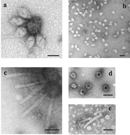
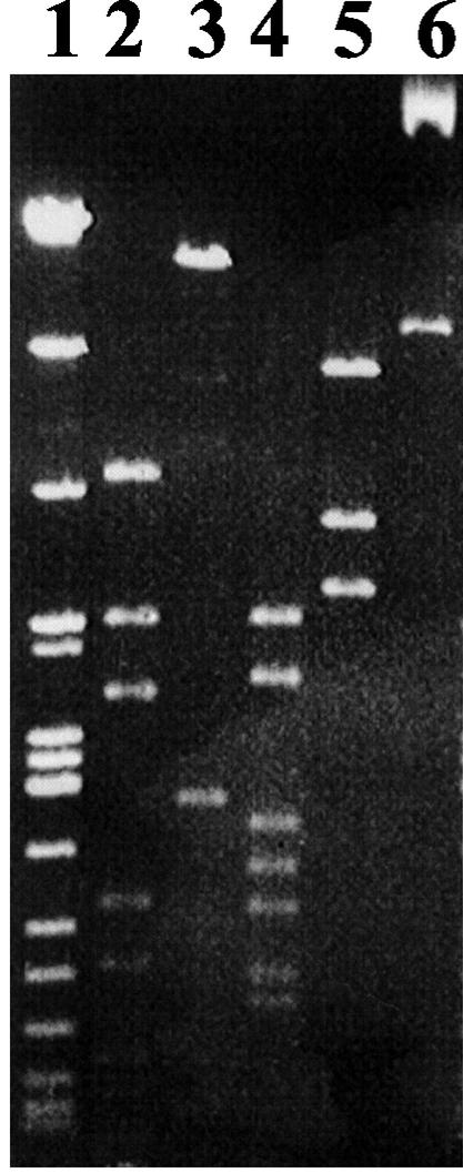
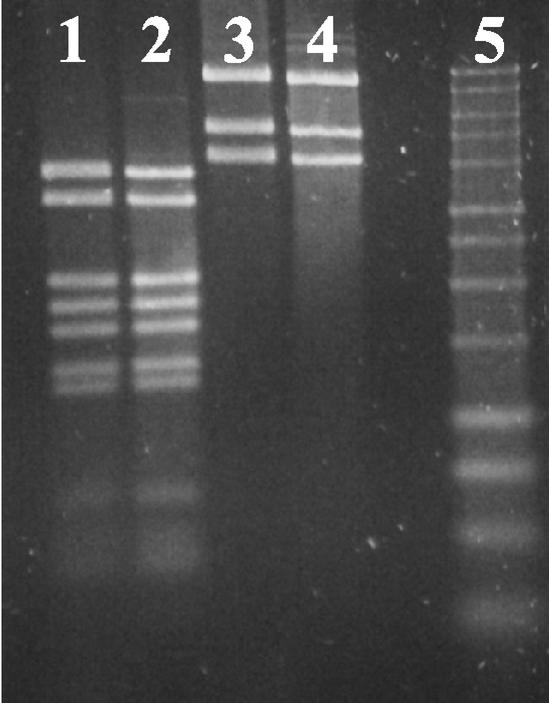
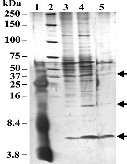
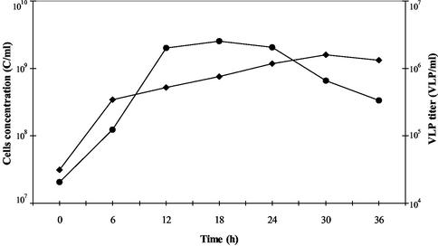
We describe the first virus-like particle of a hyperthermophilic euryarchaeote which was discovered in a strain of "Pyrococcus abyssi" previously characterized in our laboratory. This particle, named PAV1, is lemon-shaped (120 nm x 80 nm), with a short tail terminated by fibers, and resembles the virus SSV1, the type member of the Fuselloviridae, isolated from Sulfolobus shibatae. Sensitivity of the virus-like particle to organic solvents and detergents suggested that the envelope of PAV1 may contain lipids in addition to proteins. It contains a double-stranded circular DNA of 18 kb which is also present in high copy number in a free form in the host cytoplasm. No integrated form of the PAV1 genome could be detected in the host chromosome. Under standard growth conditions, the host cells continuously release PAV1 particles into the culture supernatant without spontaneous lysis, with a maximum reached in the late stationary phase. UV, gamma irradiation, treatment with mitomycin C, and various physiological stresses had no effect on PAV1 production. Screening of a large number of Thermococcales isolates did not permit to find a sensitive host. These results suggest that PAV1 persists in the host strain in a stable carrier state rather than a prophage.
Figures
References
-
- Ackerman, H.-W. 2001. Frequency of morphological phage descriptions in the year 2000. Arch. Virol. 146:843-857. - PubMed
-
- Arnold, H. P., W. Zillig, U. Ziese, I. Holz, M. Crosby, T. Utterback, J. F. Weidmann, J. K. Kristjansson, H. P. Klenk, K. E. Nelson, and C. M. Fraser. 2000. A novel Lipothrixvirus, SIFV, of the extremely thermophilic crenarchaeon Sulfolobus. Virology 267:252-266. - PubMed
-
- Arnold, H. P., U. Ziese, and W. Zillig. 2000. SNDV, a novel virus of the extremely thermophilic and acidophilic archaeon Sulfolobus. Virology 272:409-416. - PubMed
-
- Benbouzid-Rollet, N., P. Lopez-Garcia, L. Watrin, G. Erauso, D. Prieur, and P. Forterre. 1997. Isolation of new plasmids from hyperthermophilic Archaea of the order Thermococcales. Res. Microbiol. 148:1-9. - PubMed
Publication types
MeSH terms
Substances
LinkOut - more resources
Full Text Sources
Molecular Biology Databases
