. 2003 Jul;185(13):3918-25.
doi: 10.1128/JB.185.13.3918-3925.2003.

Archaeal homolog of bacterial type IV prepilin signal peptidases with broad substrate specificity

Affiliations

Archaeal homolog of bacterial type IV prepilin signal peptidases with broad substrate specificity

Sonja-Verena Albers et al. J Bacteriol. 2003 Jul.

Abstract

A large number of secretory proteins in the thermoacidophile Sulfolobus solfataricus are synthesized as a precursor with an unusual leader peptide that resembles bacterial type IV prepilin signal sequences. This set of proteins includes the flagellin subunit but also various solute binding proteins. Here we describe the identification of the S. solfataricus homolog of bacterial type IV prepilin peptidases, termed PibD. PibD is an integral membrane protein that is phylogenetically related to the bacterial enzymes. When heterologously expressed in Escherichia coli, PibD is capable of processing both the flagellin and glucose-binding protein (GlcS) precursors. Site-directed mutagenesis of the GlcS signal peptide shows that the substrate specificity of PibD is consistent with the variations found in proteins with type IV prepilin-like signal sequences of S. solfataricus. We conclude that PibD is responsible for the processing of these secretory proteins in S. solfataricus.


Figures

FIG. 1.
FIG. 1.
In vitro cleavage of glucose-binding protein (GlcS*) and flagellin (FlaB) precursors. (A) For the amino-terminal sequences of preGlcS and preFlaB, the cleavage sites are indicated by an arrow. (B) Solubilized E. coli membranes containing preGlcS* or preFlaB and S. solfataricus membranes were mixed as indicated. No cross-reaction of S. solfataricus membrane proteins with the anti-His antibody was observed (lane 2). The asterisk indicates an unspecific degradation product of preFlaB.
FIG. 2.
FIG. 2.
Identification and heterologous expression of SSO0131 (pibD). (A) Multiple alignment carried out by using Multalin (9) of the PibD primary sequence with archaeal homologs from S. tokodaii (ST2258), M. acetivorans (MA3102), A. fulgidus (AF0936), and M. maripaludis (FlaK) reveals putatively conserved residues. The sequence motifs surrounding the conserved aspartate residues that might be involved in catalysis are boxed. Residues that are 100%, greater than 80%, or greater than 60% conserved are shaded black, dark gray, and light gray, respectively. (B) Membrane topology of PibD predicted with TMHMM2 (23, 33). Aspartate residues are highlighted and those conserved in at least three sequences are highlighted with filled circles. (C) Heterologous expression of pibD in E. coli. Inner membranes isolated from cells expressing the gene (lane 1) or harboring the empty vector (lane 2) were analyzed by SDS-PAGE and detected with an antibody directed against the C-terminal hexa-His epitope tag.
FIG. 3.
FIG. 3.
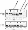
Temperature dependent in vitro cleavage of preFlaB (top panel) and preGlcS* (lower panel) by heterologously expressed PibD. Solubilized inner membranes from E. coli containing preFlaB or preGlcS* were incubated with E. coli membranes containing overexpressed PibD protein and incubated at the temperatures indicated. No processing was observed at 20°C or when membranes isolated from cells harboring the empty vector were used.
FIG. 4.
FIG. 4.
In vitro processing of preGlcS* signal peptide mutants. (A) The signal sequence of GlcS is shown at the top; the cleavage site is indicated by a gap. The mutations introduced are listed with the nomenclature used in panel B. (B) GlcS* precursors with altered signal sequences were incubated with membranes from either S. solfataricus membranes (top panel) or E. coli overexpressing recombinant pibD (lower panel). As controls, preGlcS* was incubated in absence or presence of cleavage activity, respectively (first two lanes in each panel).

References

    1. Albers, S. V., and A. J. M. Driessen. 2002. Signal peptides of secreted proteins of the archaeon Sulfolobus solfataricus: a genomic survey. Arch. Microbiol. 177:209-216. - PubMed
    1. Albers, S. V., M. G. Elferink, R. L. Charlebois, C. W. Sensen, A. J. M. Driessen, and W. N. Konings. 1999. Glucose transport in the extremely thermoacidophilic Sulfolobus solfataricus involves a high-affinity membrane-integrated binding protein. J. Bacteriol. 181:4285-4291. - PMC - PubMed
    1. Albers, S. V., W. N. Konings, and A. J. M. Driessen. 1999. A unique short signal sequence in membrane-anchored proteins of Archaea. Mol. Microbiol. 31:1595-1596. - PubMed
    1. Bardy, S. L., and K. F. Jarrell. 2002. FlaK of the archaeon Methanococcus maripaludis possesses preflagellin peptidase activity. FEMS Microbiol. Lett. 208:53-59. - PubMed
    1. Bayley, D. P., and K. F. Jarrell. 1998. Further evidence to suggest that archaeal flagella are related to bacterial type IV pili. J. Mol. Evol. 46:370-373. - PubMed

Publication types

MeSH terms

LinkOut - more resources