Comparative Study
. 2003 Jul 8;100(14):8484-9.
doi: 10.1073/pnas.0832438100. Epub 2003 Jun 18.

Conservation of genome content and virulence determinants among clinical and environmental isolates of Pseudomonas aeruginosa

Affiliations
Comparative Study

Conservation of genome content and virulence determinants among clinical and environmental isolates of Pseudomonas aeruginosa

Matthew C Wolfgang et al. Proc Natl Acad Sci U S A. .

Abstract

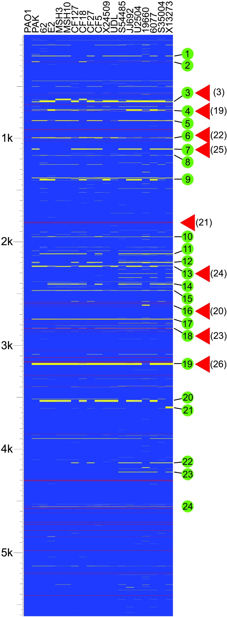
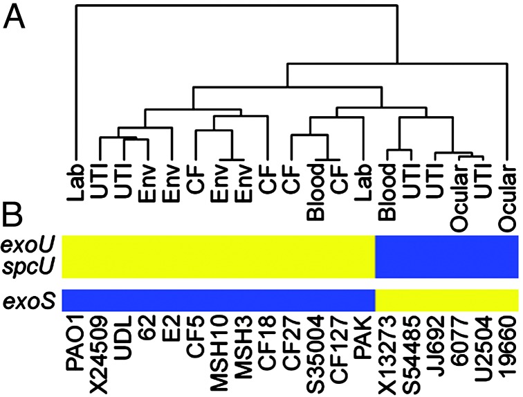
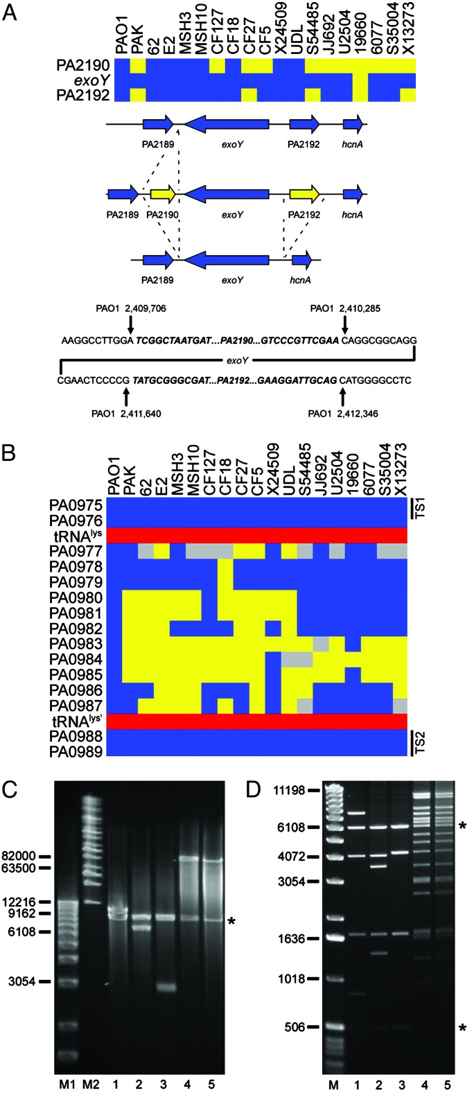
Pseudomonas aeruginosa is a ubiquitous environmental bacterium capable of causing a variety of life-threatening human infections. The genetic basis for preferential infection of certain immunocompromised patients or individuals with cystic fibrosis by P. aeruginosa is not understood. To establish whether variation in the genomic repertoire of P. aeruginosa strains can be associated with a particular type of infection, we used a whole-genome DNA microarray to determine the genome content of 18 strains isolated from the most common human infections and environmental sources. A remarkable conservation of genes including those encoding nearly all known virulence factors was observed. Phylogenetic analysis of strain-specific genes revealed no correlation between genome content and infection type. Clusters of strain-specific genes in the P. aeruginosa genome, termed variable segments, appear to be preferential sites for the integration of novel genetic material. A specialized cloning vector was developed for capture and analysis of these genomic segments. With this capture system a site associated with the strain-specific ExoU cytotoxin-encoding gene was interrogated and an 80-kb genomic island carrying exoU was identified. These studies demonstrate that P. aeruginosa strains possess a highly conserved genome that encodes genes important for survival in numerous environments and allows it to cause a variety of human infections. The acquisition of novel genetic material, such as the exoU genomic island, through horizontal gene transfer may enhance colonization and survival in different host environments.


Figures

Fig. 1.
Fig. 1.
Analysis of genome content indicates a high level of gene conservation. The diagram indicates the presence and absence of genes found in the sequenced genome of strain PAO1 in clinical and environmental isolates of P. aeruginosa as detected by microarray hybridization. Strains are indicated at the top. The source of each strain is as follows: PAO1, reference strain; PAK, laboratory strain; environmental isolates (62, E2, MSH3, MSH10); respiratory isolates from children (<24 months) with cystic fibrosis (CF127, CF18, CF27, CF5); isolates from urinary tract infections (X24509, UDL, S54485, JJ692, U2504); strains isolated from ocular infections (19660, 6077), and blood isolates (S35004, X13273). Horizontal lines represent genes. Blue indicates that a gene is present, yellow represents absence, and gray indicates that presence was indeterminate. Red lines represent the location of tRNA genes in strain PAO1. The scale represents 5,613 genes (including 5,549 ORFs and 64 tRNA-encoding genes) organized according to the PAO1 chromosome. Green circles indicate variable segments in the P. aeruginosa genome as discussed in the text. Red triangles indicate known sites of insertion of horizontally acquired genetic material. References are indicated in parentheses.
Fig. 2.
Fig. 2.
Phylogenetic analysis of P. aeruginosa isolates based on genome content indicates a limited clonal relationship but no correlation with disease. (A) Dendrogram showing the relationship between strains based on the presence and absence of genes designated as strain specific. The source of each strain is indicated. Analysis was performed by using a previously described algorithm for the hierarchical clustering of microarray hybridization data (12). (B) Correlation between a group of clonally related strains and the presence of certain strain-specific virulence genes. The presence (blue) and absence (yellow) of genes encoding ExoU, SpcU, and ExoS are indicated. Strains are arranged according to their position in the accompanying dendrogram.
Fig. 3.
Fig. 3.
Analysis of polymorphic genes and variable segments associated with type III secreted effector-encoding genes. (A) Loss of genes in the flanking region of exoY. (Top) Diagram showing the presence (blue) and absence (yellow) of exoY and flanking genes in the indicated strains. (Middle) Summary of the specific deletion/insertion events detected by PCR analysis. Not shown is the deletion of all three genes in strain 19660. (Bottom) Sequence analysis of the exoY flanking regions indicates the specific deletion/insertion junctions for strains PAK, CF127, CF27, CF5, S54485, JJ692, U2504, 6077, S35004, and X13273. Sequences in bold/italics are absent in the corresponding deletion strains indicated as yellow (Top). Coordinates of the junction sites relative to the PAO1 chromosome are given. (B) Diagram showing a variable segment of P. aeruginosa genomes associated with the acquisition of exoU. Blue boxes indicate the presence of PAO1 genes in the interrogated strains. Yellow indicates undetected genes, and gray represents genes for which no definitive determination could be made. Red boxes show the location of an intact tRNAlys gene and a partial duplication of the same gene in the PAO1 genome. Black bars indicate the location of conserved targeting sequences (TS1 and TS2) used in the construction of the capture vector. (C) Size determination of captured sequences between conserved genes PA0975 and PA0989 in five P. aeruginosa strains. DNA fragments were analyzed on a 1% agarose gel by pulsed-field gel electrophoresis using the Bio-Rad Chef Mapper System. Lane M1, 1-kb DNA ladder (Invitrogen); lane M2, Midrange I PFG Marker (New England Biolabs). Subsequent lanes contain plasmids with captured sequences from the indicated strains. Inserts were separated from the vector (indicated by *) by digestion with I-SceI. Lane 1, PAO1; lane 2, PAK; lane 3, CF127; lane 4, 6077; lane 5, JJ692. (D) Restriction fragment fingerprinting of captured sequences. Lane M, 1-kb DNA ladder. The following lanes contain NcoI digested plasmids with captured sequences from the indicated strains: 1, PAO1; 2, PAK; 3, CF127; 4, 6077; and 5, JJ692. Asterisks denote the location of vector fragments.

References

    1. Mahajan-Miklos, S., Rahme, L. G. & Ausubel, F. M. (2000) Mol. Microbiol. 37 981-988. - PubMed
    1. Bodey, G. P., Bolivar, R., Fainstein, V. & Jadeja, L. (1983) Rev. Infect. Dis. 5 279-313. - PubMed
    1. Stover, C. K., Pham, X. Q., Erwin, A. L., Mizoguchi, S. D., Warrener, P., Hickey, M. J., Brinkman, F. S., Hufnagle, W. O., Kowalik, D. J., Lagrou, M., et al. (2000) Nature 406 959-964. - PubMed
    1. Fitzgerald, J. R. & Musser, J. M. (2001) Trends Microbiol. 9 547-553. - PubMed
    1. Dziejman, M., Balon, E., Boyd, D., Fraser, C. M., Heidelberg, J. F. & Mekalanos, J. J. (2002) Proc. Natl. Acad. Sci. USA 99 1556-1561. - PMC - PubMed

Publication types

MeSH terms

Substances

LinkOut - more resources