. 2003 Sep 1;551(Pt 2):433-46.
doi: 10.1113/jphysiol.2003.039990. Epub 2003 Jun 18.

Signalling pathways involved in the sensitisation of mouse nociceptive neurones by nerve growth factor

Affiliations

Signalling pathways involved in the sensitisation of mouse nociceptive neurones by nerve growth factor

Jennifer K Bonnington et al. J Physiol. .

Abstract

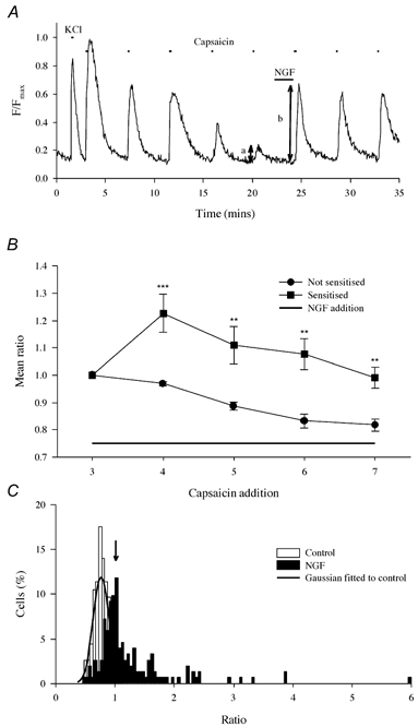
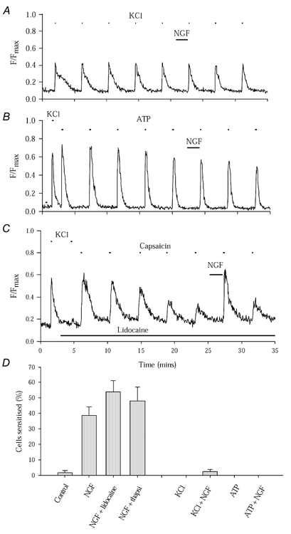
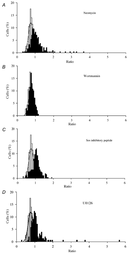
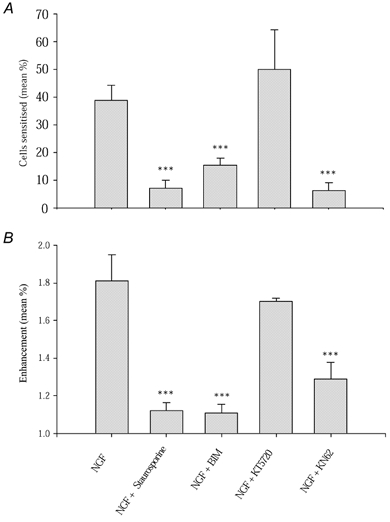
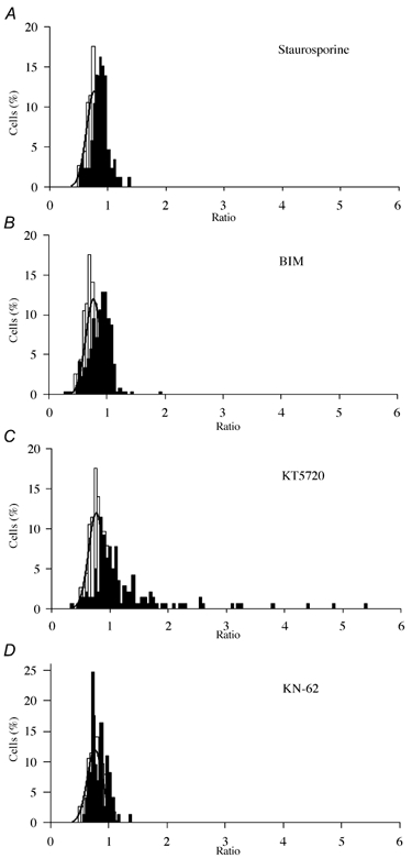
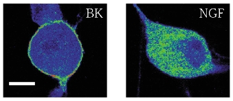
Nerve growth factor (NGF) causes a rapid sensitisation of nociceptive sensory neurones to painful thermal stimuli owing to an action on the heat and capsaicin receptor TRPV1 (formerly known as VR1). We have developed a new technique to study this rapid sensitisation of TRPV1 by monitoring the effects of NGF on the increase in intracellular calcium concentration ([Ca2+]i) following exposure to capsaicin. Brief applications of capsaicin caused a rise in [Ca2+]i, and NGF was found to enhance this rise in 37 % of capsaicin-responsive neurones within 2 min. Pathways responsible for transducing the sensitisation of TRPV1 by TrkA, the NGF receptor, were characterised by observing the effects of inhibitors of key members of NGF-activated second messenger signalling cascades. Specific inhibitors of the ras/MEK (mitogen-activated protein and extracellular signal-regulated kinases) pathway and of phospholipase C did not abolish the NGF-induced sensitisation, but wortmannin, a specific inhibitor of phosphatidylinositol-3-kinase (PI3K), totally abolished the effect of NGF. Pharmacological blockade of protein kinase C (PKC) or calcium-calmodulin-dependent protein kinase II (CaMK II) activation also prevented NGF-induced sensitisation, while blockade of protein kinase A (PKA) was without effect. These data indicate that the crucial early pathway activated by NGF involves PI3K, while PKC and CaMK II are also involved, probably at subsequent stages of the NGF-activated signalling pathway.


Figures

Figure 1
Figure 1. Enhancement of the capsaicin-induced Ca2+ increase by nerve growth factor (NGF)
A, trace illustrating a typical experimental procedure. Ca2+ increases shown in a single neurone from a coverslip containing (typically) 10–30 neurones. Neurones loaded with the Ca2+-sensitive fluorophore fluo-4 as detailed in Methods. The Ca2+ increase observed on application of KCl (25 mm, 15 s first exposure) distinguished neuronal from non-neuronal cells. Subsequent applications of capsaicin (500 nm, 15 s) caused activation of TRPV1, which was potentiated by exposure to NGF (100 ng ml−1, 2 min). NGF did not itself cause an increase in [Ca2+]i in any experiment. The ratio b/a of the Ca2+ increases before and after exposure to NGF was used as an index of enhancement. B, NGF causes a long-lasting enhancement. Neurones (ncell = 32) were separated into two groups: those in which an enhancement was (▪) or was not (•) observed in the first exposure following NGF addition (lower bar). Bars give ± s.e.m. Significance levels (two-tailed t test) are: **P < 10−2; ***P < 10−3. (NB in this experiment a continuous NGF exposure followed the third capsaicin application while in all others a 2 min exposure was given between the fifth and sixth capsaicin applications). C, collected ratio values obtained from experiments as in A. Open bars give ratios obtained without exposure to NGF (ncell = 112, nexp = 8). The distribution was well fitted by a Gaussian function with mean of 0.76, s.d. of 0.134 and upper 95 % two-tailed confidence limit of 1.023 (arrow). Filled bars give ratios following 2 min exposure to NGF (100 ng ml−1; ncell = 152, nexp = 26). Following NGF exposure 38.74 ± 5.61 % of ratios exceeded the 95 % confidence limit, and the mean of these ratio values was 1.81 ± 0.14.
Figure 2
Figure 2. NGF enhances Ca2+ entry through TRPV1 but not via other routes
A, NGF has no effect on the Ca2+ increase evoked by KCl (25 mm, 9 s). B, NGF has no effect on the Ca2+ increase evoked by ATP (100 μm, 15 s). Neurones incubated with thapsigargin (10 μm) for 20 min prior to experiments; the Ca2+ increase evoked by thapsigargin lasted 5–7 min, showing that stores were completely emptied with a 20 min pre-exposure. C, enhancement of TRPV1 function by NGF is observed following complete block of voltage-sensitive ion channels by lidocaine (lignocaine; 2 mm, black bar). KCl-evoked Ca2+ increase (see two applications at start) is completely abolished by lidocaine but enhancement of capsaicin-evoked [Ca2+]i increase by NGF is still observed. D, collected results of experiments similar to those in A, B and C, together with experiment similar to C in which thapsigargin (10 μm, applied 20 min prior to experiment) was used to empty subcellular Ca2+ stores. Bars give ± s.e.m. Numbers of cells and separate experiments as follows: control, ncell = 112, nexp = 8; NGF alone, ncell = 152, nexp = 26; NGF + lidocaine, ncell = 60, nexp = 8; NGF + thapsigargin, ncell = 71, nexp = 16; KCl, ncell = 88, nexp = 7; KCl + NGF, ncell = 144, nexp = 7; ATP, ncell = 23, nexp = 9; ATP + NGF, ncell = 63, nexp = 15.
Figure 4
Figure 4. Sensitisation of TRPV1 by NGF following inhibition of PLC, PI3K, ras and MEK
A, the proportion of neurones sensitised in the presence of 10 μm neomycin, 20 nm wortmannin, 10 μm Sos-inhibitory peptide and 10 μm U0126, to inhibit PLC, PI3K, ras and MEK activation respectively. B, mean ratio values of sensitised neurones. Error bars show means ± s.e.m. Significance levels: *P < 5 %; **P < 1 %; ***P < 0.1 %.
Figure 3
Figure 3. The effects of inhibition of PLC, PI3K, ras, and MEK1 and 2 on NGF-induced sensitisation of TRPV1
Ratio distributions as in Fig. 1C are shown for NGF (100 ng ml−1) plus the following treatments, compared with control ratio distribution in absence of NGF (fitted with Gaussian function as in Fig. 1C): A, 10 μm neomycin; B, 20 nm wortmannin; C, 10 μm Sos-inhibitory peptide; D, 10 μm U0126. Inhibitors were applied 5 min before experiment and were included in all solutions. Numbers of cells and separate experiments as follows: neomycin, ncell = 240, nexp = 33; wortmannin, ncell = 139, nexp = 20; Sos-inhibitory peptide, ncell = 259, nexp = 20; U0126, ncell = 155, nexp = 8.
Figure 6
Figure 6. Sensitisation of TRPV1 by NCF following inhibition of PKC, PKA and CaMK II
A, the proportion of neurones sensitised by NGF in the presence of 200 nm staurosporine, 500 nm BIM to inhibit PKC, 200 nm KT5720 to inhibit PKA and 1 μm KN-62 to block CaMK II activity. B, mean ratio values of sensitised neurones. Error bars show means ± s.e.m. Significance levels: ***P < 0.1 %.
Figure 5
Figure 5. The effect of inhibition of PKC, PKA and CaMK II on NGF-induced sensitisation of TRPV1
Ratio distributions as in Fig. 1C shown for NGF (100 ng ml−1) plus the following treatments, compared with control ratio distribution in absence of NGF (fitted with Gaussian function as in Fig. 1C): A, 200 nm staurosporine; B, 500 nm BIM; C, 200 nm KT5720; D, 1 μm KN-62. Inhibitors were pre-applied for 5 min and were included in all solutions throughout the experiment. Numbers of cells and separate experiments as follows: staurosporine, ncell = 86, nexp = 7; BIM, ncell = 264, nexp = 15; KT5720, ncell = 141, nexp = 7; KN-62, ncell = 73, nexp = 8.
Figure 7
Figure 7. Cellular location of PKCε following exposure to bradykinin (left, 1 μm for 30 s) and NGF (100 ng ml−1, 2 min)
Typical images of neurones fixed and imaged in a confocal microscope as described in Methods. False-colour images with blue indicating low fluorescence and red high fluorescence. Scale bar: 10 μm.

References

    1. Aley KO, Martin A, McMahon T, Mok J, Levine JD, Messing RO. Nociceptor sensitisation by extracellular signal-regulated kinases. J Neurosci. 2001;21:6933–6939. - PMC - PubMed
    1. Bergmann I, Reiter R, Toyka KV, Koltzenburg M. Nerve growth factor evokes hyperalgesia in mice lacking the low-affinity neurotrophin receptor p75. Neurosci Lett. 1998;255:87–90. - PubMed
    1. Bhave G, Zhu W, Wang H, Brasier DJ, Oxford GS, Gereau RW. cAMP-dependent protein kinase regulates desensitisation of the capsaicin receptor (VR1) by direct phosphorylation. Neuron. 2002;35:721–731. - PubMed
    1. Broad LM, Cannon TR, Taylor CW. A non-capacitative pathway activated by arachidonic acid is the major Ca2+ entry mechanism in rat A7r5 smooth muscle cells stimulated with low concentrations of vasopressin. J Physiol. 1999;517:121–134. - PMC - PubMed
    1. Bron R, Klesse LJ, Shah K, Parada LF, Winter J. Activation of Ras is necessary and sufficient for upregulation of vanilloid receptor type 1 in sensory neurons by neurotrophic factors. Mol Cell Neurosci. 2003;22:118–132. - PubMed

Publication types

MeSH terms

LinkOut - more resources