. 2003 Sep 15;551(Pt 3):751-63.
doi: 10.1113/jphysiol.2003.040014. Epub 2003 Jun 18.

Voltage-gated K+ channels in rat small cerebral arteries: molecular identity of the functional channels

Affiliations

Voltage-gated K+ channels in rat small cerebral arteries: molecular identity of the functional channels

Sulayma Albarwani et al. J Physiol. .

Abstract

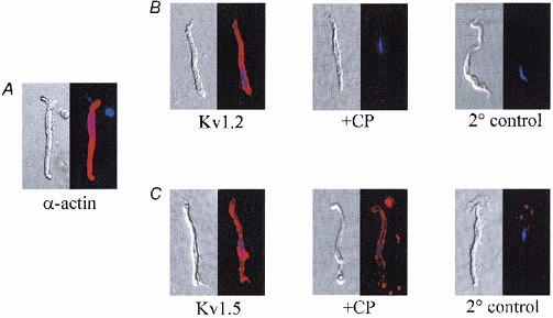
Voltage-gated potassium (KV) channels represent an important dilator influence in the cerebral circulation, but the composition of these tetrameric ion channels remains unclear. The goals of the present study were to evaluate the contribution of KV1 family channels to the resting membrane potential and diameter of small rat cerebral arteries, and to identify the alpha-subunit composition of these channels using patch-clamp, molecular and immunological techniques. Initial studies indicated that 1 micromol l(-1) correolide (COR), a specific antagonist of KV1 channels, depolarized vascular smooth muscle cells (VSMCs) in pressurized (60 mmHg) cerebral arteries from -55 +/- 1 mV to -34 +/- 1 mV, and reduced the resting diameter from 152 +/- 15 microm to 103 +/- 20 microm. In patch clamped VSMCs from these arteries, COR-sensitive KV1 current accounted for 65 % of total outward KV current and was observed at physiological membrane potentials. RT-PCR identified mRNA encoding each of the six classical KV1 alpha-subunits, KV1.1-1.6, in rat cerebral arteries. However, only the KV1.2 and 1.5 proteins were detected by Western blot. The expression of these proteins in VSMCs was confirmed by immunocytochemistry and co-immunoprecipitation of KV1.2 and 1.5 from VSMC membranes suggested KV1.2/1.5 channel assembly. Subsequently, the pharmacological and voltage-sensitive properties of KV1 current in VSMCs were found to be consistent with a predominant expression of KV1.2/1.5 heterotetrameric channels. The findings of this study suggest that KV1.2/1.5 heterotetramers are preferentially expressed in rat cerebral VSMCs, and that these channels contribute to the resting membrane potential and diameter of rat small cerebral arteries.


Figures

Figure 1
Figure 1. Effects of correolide (COR) and 4-aminopyridine (4-AP) on the resting Vm and diameter of rat small cerebral arteries
A, microelectrode recordings of Vm in a rat middle cerebral artery indicate a resting Vm level of about −57 mV. B, the addition of 1 μM correolide (COR) induced a pronounced depolarization to about −34 mV (top trace). COR induced action potentials in another artery (bottom trace). C, the final addition of 1 mM 4-AP did not induce further depolarization. D and E, average Vm and diameter values for cerebral arteries from six rats exposed to 1 μM COR, and subsequently, exposed to both 1 μM COR and 1 mM 4-AP. Data are expressed as means ±s.e.m. (n = 6). * Significant difference (P < 0.05) between control and experimental values.
Figure 2
Figure 2. Verification of K+ selectivity of outward current
A, outward currents in rat cerebral VSMCs elicited by progressive 8 mV depolarizing steps from a holding potential of −70 mV to +58 mV. B, tail currents were generated by hyperpolarizing steps to between −100 and −4 mV (8 mV increments), following an initial activating pre-pulse from −70 to +58 mV. C-E, reversal potentials of VSMCs dialysed with pipette solution containing 145 mM K+, and bathed in external solution containing 4, 20 and 70 mM K+ (n = 4–6 cells averaged from 4–5 rats).
Figure 3
Figure 3. Effects of correolide (COR) and 4-aminopyridine (4-AP) on voltage-gated K+ currents in rat cerebral VSMCs
A, whole-cell K+ currents elicited in physiological K+ gradients were dose-dependently blocked by 0.1 and 1 μM correolide (COR), a selective blocker of KV1 channels. The further addition of 4-AP had little effect. Cell capacitance was 15 pF. B-D, averaged I-V relations demonstrating the effect of 0.1 μM COR, 1 μM COR, and the combined effect of 1 μM COR and 1 mM 4-AP on KV currents. Data are expressed as means ±s.e.m. (n = 6–9). * Significant difference (P < 0.05) between control and experimental values.
Figure 4
Figure 4. Detection of amplified products corresponding to KV1.1–1.6 mRNA in rat brain and cerebral arteries
A, RT-PCR screening detected mRNA encoding KV1.1–1.6 α-subunits in lanes loaded with amplified cDNA originating from rat brain (Br, left lanes) or rat middle cerebral arteries (C, right lanes). Detection of each product was verified in cDNA reverse transcribed from 7–12 different RNA isolations. Each isolation was prepared from tissues of two rats. The brain was used as a positive control. Expected product sizes (bp) were: 284 (1.1), 374 (1.2), 581 (1.3), 643 (1.4), 1111 (1.5), 578 (1.6). B, screening for genomic contamination of cDNA using primers for smooth muscle-specific α-actin that included three introns in the amplified region. Amplification reactions including cDNA (70 nmol) resulted in a 637 bp product consistent with the mRNA template, whereas reactions including DNA (70 nmol) resulted in the predicted 1506 bp product amplified from the intron-containing region. Control reactions included amplifications without cDNA or without DNA. Results were verified in at least three different preparations. Each preparation was obtained from tissues of two rats.
Figure 5
Figure 5. Screening by Western blots for KV1.1–1.6 proteins in rat brain and cerebral arteries
A, Western blots showed immunoreactive bands corresponding to KV1.1, 1.3, 1.4 and 1.6 α-subunits in rat brain (Br, left lanes), but not in rat cerebral arteries (C, right lanes). The apparent molecular mass values of the brain proteins were approximately: 60 kDa (1.1), 90 kDa (1.3), 85 kDa (1.4) and 60 kDa (1.6). B, KV1.2 was detected in rat brain (Br) and cerebral arteries (C) at a molecular mass of ˜80 kDa. The immunoreactive band in the arterial proteins was eliminated by an antigenic competing peptide (+CP). C, brain (Br) or cerebral artery (C) membrane proteins were prepared in control buffer or were enzymatically treated with PNGase F (+P) before analysis by electrophoresis. Treatment with PNGase F reduced the apparent molecular mass of KV1.2 in both types of tissue, implying N-glycosylation of the mature protein. D, the KV1.5 α-subunit was detected as a ˜75 kDa band in cerebral arteries (C), but was absent in the brain. The immunoreactive band was eliminated by a competing peptide (+CP). Western results were verified in 3 (KV1.1, 1.3, 1.4, 1.6), 16 (KV1.2), 4 (KV1.2 PNGase F) and 6 (KV1.5) different protein preparations isolated from 4–10 rats.
Figure 6
Figure 6. Normarski and corresponding fluorescent images of freshly isolated rat cerebral VSMCs
A, expression of smooth muscle-specific α-actin. Cells were visualized using Alexa Fluor 594-conjugated antibody (in red). Nuclei were labelled with DAPI (in blue). B, left to right, VSMCs labelled with anti-KV1.2 (Kv1.2), after adsorption of anti-KV1.2 with a competing peptide (+CP) or after incubation with secondary antibody only (2 ° control). C, left to right, VSMCs labelled with anti-KV1.5 (Kv1.5), after adsorption of anti-KV1.5 by a competing peptide (+CP), and after incubation with secondary antibody only (2 ° control). Within each panel, fluorescent images were acquired from VSMCs exposed for equivalent times. Results were verified in 3 (α-actin), 9 (Kv1.2) and 5 (Kv1.5) cells from different preparations.
Figure 7
Figure 7. Co-immunoprecipitation of KV1.2 and 1.5 from rat cerebral arteries
A, stably expressed KV1.5 in HEK cell membranes (1 μg) detected by anti-KV1.5 blot as a positive control. B, anti-KV1.5 immunoprecipitate from cerebral arterial membranes (200 μg) blotted with anti-KV1.5 revealed a 75 kDa band, indicating successful immunoprecipitation of KV1.5. C, stably expressed KV1.2 in HEK cell membranes (1 μg) detected by anti-KV1.2 blot as a positive control. D, native KV1.2 in cerebral arterial membranes (8 μg) detected by anti-KV1.2 blot as an ˜80 kDa band. E and F, anti-KV1.5 immunoprecipitate from 50 μg and 30 μg of two different cerebral arterial membrane preparations. An anti-KV1.2 blot revealed the same ˜80 kDa band in both arterial preparations, indicating successful co-immunoprecipitation of KV1.2 and 1.5. G, preadsorption of anti-KV1.2 with 1 μM of its antigenic competing peptide (+CP) abolished the 80 kDa-immunoreactive band detected in anti-KV1.5 immunoprecipitate. Anti-KV1.5 immunoprecipitate was from 30 μg of the same pool of arterial proteins as used in F. Results using cerebral arteries were verified in a minimum of four different protein preparations isolated from ˜60 rats.
Figure 8
Figure 8. KV1 current is insensitive to α-dendrotoxin and margatoxin in rat cerebral VSMCs
A, potassium currents generated by 8 mV depolarizing steps from −70 to +58 mV were only slightly reduced by α-dendrotoxin (α-DTX, 100 nM) and margatoxin (MgTX, 100 nM). Both drugs block KV1.2 homotetramers, but not KV1.5 homotetramers or KV1.2/1.5 heterotetrameric channels. The addition of 1 μM correolide (COR) reduced the same current. B, I-V relations averaged from six cells demonstrate the relative toxin-insensitivity of K+ currents in cerebral VSMCs, but the sensitivity of the same currents to block by COR. Data are expressed as means ±s.e.m. (n = 6). * Significant difference (P < 0.05) between control and experimental values at the same voltage.
Figure 9
Figure 9. Analysis of the voltage sensitivity of correolide (COR)-sensitive KV1 current
A, pulse protocol used to assess the effect of 1 μM correolide (COR) on K+ channel activation in symmetrical K+ solutions. The two superimposed tail currents evoked by a voltage step from −70 to +58 mV demonstrate COR-sensitive tail currents. B, the effect of 1 μM COR on steady-state activation (n = 6). Values were provided by tail current analysis. C, pulse protocol used to assess the effect of 1 μM COR on inactivation in physiological K+ gradients. Superimposed traces were evoked by inactivating pre-pulses of −86, −14 and +18 mV, followed by a depolarizing step to +58 mV after a 5 ms delay at −70 mV to evaluate available current. D, the effect of 1 μM COR on steady-state inactivation (n = 7). Values were provided by experiments detailed in C. E, activation and inactivation relationships for COR-sensitive current. Values were obtained by subtracting COR-insensitive current from total K+ current, and were fitted with a Boltzmann function. The V0.5 value for activation was −1 mV and k was 14, whereas the V0.5 value for inactivation was −37 mV and k was 12. Data are expressed as means ±s.e.m. (n = 6–7). * Significant difference (P < 0.05) between control and experimental values at the same voltage.

References

    1. Aiello EA, Walsh MP, Cole WC. Phosphorylation by protein kinase A enhances delayed rectifier K+ current in rabbit vascular smooth muscle cells. Am J Physiol. 1995;268:H926–934. - PubMed
    1. Archer SL, Souil E, Dinh-Xuan AT, Schremmer B, Mercier J-C, El Yaagoubi A, Nguyen-Huu L, Reeve HL, Hampl V. Molecular identification of the role of voltage-gated K+ channels, Kv1. 5 and Kv2.1, in hypoxic pulmonary vasoconstriction and control of resting membrane potential in rat pulmonary artery myocytes. J Clin Invest. 1998;101:2319–2330. - PMC - PubMed
    1. Attali B, Lesage F, Ziliani P, Guillemare E, Honore E, Waldmann R, Hugnot J-P, Mattel M-G, Lazdunski M, Barhanin J. Multiple mRNA isoforms encoding the mouse cardiac Kv1–5 delayed rectifier K+ channel. J Biol Chem. 1993;268:24283–24289. - PubMed
    1. Beckh S, Pongs O. Members of the RCK potassium channel family are differentially expressed in the rat nervous system. EMBO J. 1990;9:777–782. - PMC - PubMed
    1. Cheong A, Dedman AM, Beech DJ. Expression and function of native potassium channel (KVα1) subunits in terminal arterioles of rabbits. J Physiol. 2001a;534:691–700. - PMC - PubMed

Publication types

MeSH terms

LinkOut - more resources