. 2003 Sep 1;551(Pt 2):455-66.
doi: 10.1113/jphysiol.2003.042903. Epub 2003 Jun 19.

Distribution and regulation of expression of serum- and glucocorticoid-induced kinase-1 in the rat kidney

Affiliations

Distribution and regulation of expression of serum- and glucocorticoid-induced kinase-1 in the rat kidney

D Alvarez de la Rosa et al. J Physiol. .

Abstract

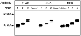
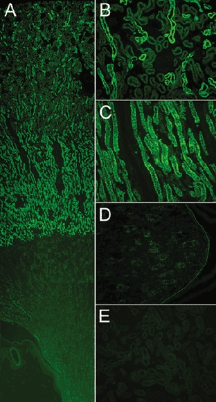
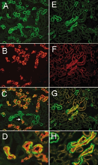
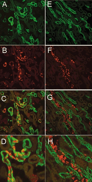
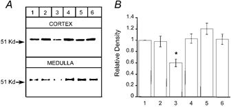
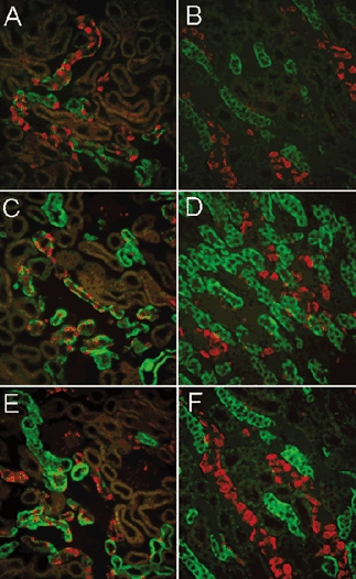
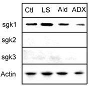
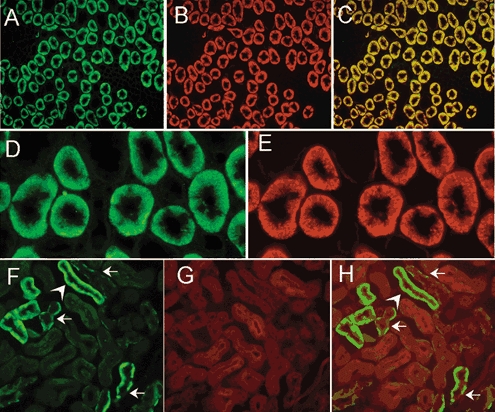
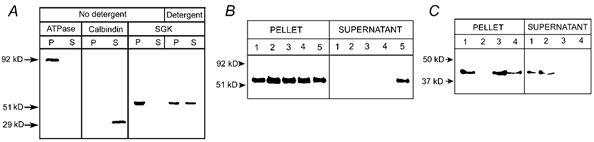
The serum- and glucocorticoid-induced kinase-1 (sgk1) increases the activity of a number of epithelial ion channels and transporters. The present study examines the distribution and subcellular localization of sgk1 protein in the rat kidney and the regulation of levels of expression induced by steroids. The results indicate that the kidney expresses predominantly the sgk1 isoform with a distribution restricted to the thick ascending limb of Henle, distal convoluted, connecting and cortical collecting tubules. Within cells, sgk1 strongly associates with the microsomal fraction of homogenates and it colocalizes with the Na+,K+-ATPase to the basolateral membrane. Analysis of the levels of expression of sgk1 by Western blotting and immunohistochemistry indicates constitutive high expression under basal conditions. Approximately half of the basal level is maintained by glucocorticoids whereas physiological fluctuations of aldosterone produce minor changes in sgk1 abundance in adrenal-intact animals. These results do not support the notion that physiological changes of aldosterone concentration turn the expression of sgk1 'on and off' in the mammalian kidney. Additionally, localization of sgk1 to the basolateral membrane indicates that the effects mediated by sgk1 do not require a direct interaction with the ion channels and transporters whose activity is modulated, since most of these proteins are located in the apical membrane of renal epithelial cells.


Figures

Figure 1
Figure 1. Specificity of the anti-sgk antibody and identification of sgk isoforms expressed in the kidney
Western blots of proteins extracted from HEK 293 cells transfected with sgk1, sgk2 and sgk3 cDNAs, control (non-transfected cells), and renal tissue examined with anti-FLAG monoclonal and anti-sgk antibodies. The lane labelled kidney contains 5-fold more protein than the lanes loaded with HEK 293 cell lysates. The arrows on the left indicate molecular masses of standards.
Figure 2
Figure 2. Distribution of sgk1 in kidneys from normal rats examined by immunohistochemistry with anti-sgk antibody
A, low magnification of a sagittal view of the kidney shows cortex, medulla and papilla. B, higher magnification of cortex; immunoreactivity is detected in distal tubules but not in proximal tubules or glomeruli. C, outer medulla; fluorescent signal localizes to arrays of tubules formed by tall cells, consistent with the thick ascending limb (TAL). There is a marked and sharp decrease in immunoreactivity in the transition from outer to inner medulla. D, papilla. A line was drawn to visualize the limit of the papilla. E, competition of primary antibody with the cognate peptide eliminates fluorescent signal from the renal cortex.
Figure 3
Figure 3. Double-labelling of sgk1 and calbidin, and sgk1 and NKCC2
Left-hand panels show renal cortex sections labelled for sgk (green) and calbindin (red). A, sgk. B, calbindin monoclonal antibody as a marker for the distal convoluted tubule (DCT) and connective tubule (CT). C, overlay of A and B shows that all tubules expressing calbindin are also positive for sgk1. The arrow points to the transition of a tubule where immunoreactivity to calbindin becomes positive. D, higher magnification of C shows tubules with sgk labelling more intense toward the basolateral membrane whereas the signal of calbindin is more intense over the apical membrane. Right -hand panels show cortex sections double-labelled for sgk1 (green) and sodium-potassium-two chloride cotransporter monoclonal antibody (NKCC2) (red). E, sgk. F, NKCC2 monoclonal antibody. Staining is restricted to the apical membrane of a few tubules that correspond to cortical segments of TAL. G, overlay of E and F shows that sgk1 is expressed in all cortical tubules positive for NKCC2. H, higher magnification of G to show tubules with apical NKCC2 and basolateral sgk1 staining.
Figure 4
Figure 4. Double-labelling of sgk1 and AE1
The left and right panels show images from renal cortex and outer medulla, respectively. A, cortex stained with sgk. B, AE1 shows staining of basolateral membranes of intercalated cells in CCT. C, overlay of A and B shows colocalization of sgk1 and AE1 in CNT and CCT. D, larger magnification of a CCT with intercalated cells stained with AE1 and principal cells with sgk1. Outer medulla stained with sgk1 (E), AE1 (F), overlay of E and F (G). H, higher magnification of G shows that sgk1 is not expressed in MCT.
Figure 6
Figure 6. Levels of expression of sgk1 analysed by Western blotting
A, representative example of a Western blot. Equal amounts of proteins from renal cortex or medulla of rats subjected to various treatments to alter glucocorticoid and mineralocorticoid concentrations were examined by Western blotting with sgk antibody. B, quantification of the intensity of the sgk1 signal by scanning densitometry of gels (n = 4). Values were normalized to the intensity of the bands from normal rats. Lanes correspond to: (1) control rats, (2) low-salt diet, (3) adrenalectomy, (4) adrenalectomy and dexamethasone infusion by minipump, (5) single dose of aldosterone and (6) spironolactone, 3 h prior to harvesting kidneys. Values are reported as means ±s.d.* Significant statistical difference (Students's t test, P < 0.001).
Figure 5
Figure 5. Distribution and levels of sgk1 expression in animals with various levels of steroids
Left and right columns correspond to sections from cortex and outer medulla, respectively. All panels show double-labelling of sgk1 (green) and AE1 (red). A and B, bilateral adrenalectomy with high-salt diet; C and D, bilateral adrenalectomy with dexamethasone infusion by minipump. E and F, low-salt diet for 1 week. Immunoreactivity is reduced by adrenalectomy but levels return to baseline values with administration of dexamethasone. The reduction in sgk1 expression is not selective to the CCT but also is seen in the TAL. Sodium depletion did not increase the signal of sgk1.
Figure 7
Figure 7. Northern blot analysis of sgk1, sgk2 and sgk3 mRNA from kidney
Equal amounts of total RNA (20 μg) extracted from kidneys of treated animals were analysed by Northern blotting using radiolabelled probes for sgk1, sgk2 and sgk3. For accurate quantification of the amount of RNA loaded, each blot was hybridized with an actin probe. All membranes were exposed to X-ray film for 12 h. Signals corresponding to sgk1 mRNA were the only signals detected. The levels changed according to the treatments. Ctl, control; LS, low-salt diet for 1 week; Ald, single injection of aldosterone; ADX, bilateral adrenalectomy.
Figure 8
Figure 8. Double-staining of kidney sections for sgk1, the Na+,K+-ATPase and actin
Staining of a transverse section of the outer medulla for sgk1 (A), the α subunit of the Na+,K+-ATPase (B), and overlay of these two images (C) showing colocalization of the two proteins. Higher magnification of the outer medulla stained for sgk1 (D), and the α subunit of the Na+,K+-ATPase (E) are also shown. Deep infolds of the basolateral membrane are delineated by the two antibodies whereas no fluorescent signal is apparent in the apical membrane. F, section of cortex stained with anti-sgk1. Arrows indicate the basolateral localization of the signal. These tubules correspond to CCT because the staining is restricted to principal cells and absent in intercalated cells. Actin labelled with DNase I conjugated with Texas red (G) distributes over the whole cytoplasm with enhancement of apical microvilli. Overlay shows sgk1 in the basolateral membrane but absent from the apical membrane (H).
Figure 9
Figure 9. sgk1 associates with the membrane fraction of kidney homogenates
A, kidneys from normal rats were homogenized in the absence of detergent or in the presence of 1 % Triton X-100. The homogenates were centrifuged at 250 000 g for 30 min and equal amounts of proteins from pellet and supernatant were examined by Western blotting with antibodies for the α subunit of Na+,K+-ATPase, calbindin and sgk1. Na+,K+-ATPase (ATPase) and sgk1 were detected only in the pellet of non-detergent homogenates whereas calbindin was detected in the supernatant. After treatment of the pellet with 1 % Triton X-100, sgk1 was partially released to the supernatant. B, the pellet obtained after the first centrifugation (250 000 g for 30 min) was resuspended in 200 μl of: (1) 0.3 m sucrose, (2) 0.1 m Na2CO3, pH 11.5, (3) 0.5 m NaCl, (4) 2 m urea or (5) 1 % Triton X-100, and centrifuged again at 250 000 g for 30 min. Equal amounts of proteins from the pellets and 30 μl from the supernatant were loaded on a gel and analysed by Western blotting with anti-sgk1 antibody. C, Western blot similar to that shown in B, but probed with a monoclonal anti-actin antibody. Arrows indicate migration of molecular mass markers.

References

    1. Alliston TN, Maiyar AC, Buse P, Firestone GL, Richards JS. Follicle stimulating hormone-regulated expression of serum/glucocorticoid-inducible kinase in rat ovarian granulosa cells: a functional role for the Sp1 family in promoter activity. Mol Endocrinol. 1997;11:1934–1949. - PubMed
    1. Alper SL, Stuart-Tilley AK, Biemesderfer D, Shmukler BE, Brown D. Immunolocalization of AE2 anion exchanger in rat kidney. Am J Physiol. 1997;274:F601–614. - PubMed
    1. Alvarez de la Rosa D, Canessa CM. Role of SGK in hormonal regulation of the epithelial sodium channel in A6 cells. Am J Physiol Cell Physiol. 2003;284:C404–414. - PubMed
    1. Alvarez de la Rosa D, Zhang P, Naray-Fejes-Toth A, Canessa CM. The serum and glucocorticoid kinase sgk increases the abundance of epithelial sodium channels in the plasma membrane of Xenopus oocytes. J Biol Chem. 1999;274:37834–37839. - PubMed
    1. Bhargava A, Fullerton MJ, Myles K, Purdy TM, Funder JW, Pearce D, Cole TJ. The serum- and glucocorticoid-induced kinase is a physiological mediator of aldosterone action. Endocrinology. 2001;142:1587–1594. - PubMed

Publication types

MeSH terms

LinkOut - more resources