Review
. 2003;5(4):208-15.
doi: 10.1186/bcr615. Epub 2003 Jun 3.

Host microenvironment in breast cancer development: epithelial-cell-stromal-cell interactions and steroid hormone action in normal and cancerous mammary gland

Affiliations
Review

Host microenvironment in breast cancer development: epithelial-cell-stromal-cell interactions and steroid hormone action in normal and cancerous mammary gland

Sandra Z Haslam et al. Breast Cancer Res. 2003.

Abstract

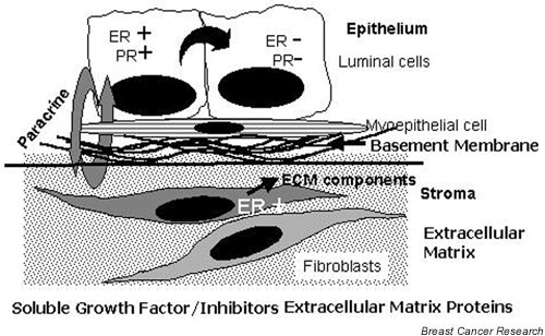
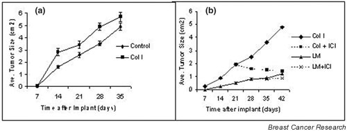
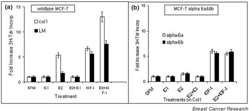
Mammary epithelial cells comprise the functional component of the normal gland and are the major target for carcinogenesis in mammary cancer. However, the stromal compartment of the normal gland and of tumors plays an important role in directing proliferative and functional changes in the epithelium. In vivo and in vitro studies of the murine mammary gland have provided insights into novel stroma-dependent mechanisms by which estrogen and progesterone action in the epithelium can be modulated by hepatocyte growth factor (HGF) and the extracellular matrix proteins, collagen type I, fibronectin and laminin. In vitro and in vivo studies of estrogen receptor positive, estrogen-responsive human breast cancer cells have also demonstrated that estrogen responsiveness of tumor cells can also be modulated by extracellular matrix proteins, collagen type I and laminin.


Figures

Figure 1
Figure 1
Model of epithelial-cell–stromal-cell interactions. ECM, extracellular matrix; ER, estrogen receptor; PR, progesterone receptor.
Figure 2
Figure 2
Response of murine mammary epithelial cells to co-culture with mammary fibroblasts and to fibroblast conditioned medium. (a) Proliferation of epithelial cells cocultured with mammary fibroblasts or in the presence of conditioned medium obtained from mammary fibroblasts. Murine mammary epithelial cells were suspended in collagen type I gels and cultured alone in basal medium (EPI), over mammary fibroblasts in basal medium (CO-CULT), or in the presence of fibroblast-conditioned medium (FCM). 3H-thymidine incorporation into DNA was assayed after 3 days of culture. *P = 0.01 that proliferation is greater under coculture condition and in the presence of FCM. (b) Phase-contrast photomicrographs (i–iv) and histological sections (v,vi) showing the tubular/ductal structure of organoids of epithelial cells in collagen-gel cell culture. Mammary epithelial cells were cultured alone in basal medium, cocultured with mammary fibroblasts in basal medium (CO-CULTURE), cultured alone in the presence of fibroblast-conditioned medium (FCM), or cultured in the presence of 50 ng/ml HGF (HGF) for 3 days. ×100 (i–iv), ×400 (v,vi). (c) Effect of FCM and estrogen on proliferation of epithelial cells. Mammary epithelial cells were cultured alone in collagen type I in basal medium, in the presence of 20 nM E2, in FCM, or in FCM obtained from fibroblasts cultured in the presence of 20 nM estradiol (E2FCM); to block any effect of estradiol in the epithelial cells, 200 nM of the antiestrogen, ICI 182,780, was added to epithelial cells at the same time as E2FCM was added. In the presence of BM (i) only solid spheres were observed. Coculture with fibroblasts (ii), or treatment with FCM (iii), or HGF (iv) produced organoids with a tubulo/ductal morphology. Organoids cultured in the presence of EGF or IGF-1 (d) produced a flattened, sheet-like morphology with few or no tubules. *P = 0.01 that proliferation in the presence of FCM was greater than in basal medium or in the presence of E2. **P = 0.05 that proliferation in the presence of E2FCM was greater than with all other treatments. (d) Morphological response of mammary epithelial cells to EGF (50 ng/ml) or IGF-1 (100 ng/ml). Phase-contrast photomicrographs of epithelial cells were taken on day 3; ×100. BM, basal medium; cpm, counts per minute; E2, 17β-estradiol; EGF, epidermal growth factor; FCM, fibroblast-conditioned medium; HGF, hepatocyte growth factor; IGF-I, insulin-like growth factor I; Tdr, thymidine.
Figure 3
Figure 3
Effect of R5020 plus HGF on proliferation of epithelial cells. Murine mammary epithelial cells were suspended in collagen type I gels and cultured in (a) HGF alone (HGF, 50 ng/ml) or with HGF in combination with E2 (10 nM), R5020 (20 nM) or E2+R5020 (10 nM+20 nM) or (b) in FCM with or without R5020 or E2+R5020. 3H-thymidine incorporation into DNA was assayed after 3 days of culture. The data are expressed for suspensions in basal medium as 3H-thymidine incorporated per well and for HGF- and FCM-treated groups as fold increase over basal-medium control. *P = 0.05 that proliferation is greater in HGF+R5020 group than in HGF or HGF+E2. ** P = 0.01 that the fold increase in proliferation in suspensions in HGF+ E2+R5020 and FCM+ E2+R5020 is greater than in all other groups within the same experiment. (c) Phase-contrast photomicrographs of epithelial cell organoid morphology in collagen gel cell culture after 3 days in basal medium containing R5020, RU486, HGF, R5020+HGF, RU486+R5020, or RU486+R5020+HGF. ×100. Note appearance of lumens (L) and alveolar buds (AB) in R5020 and R5020+HGF-treated cultures, respectively, and long tubules (T) in HGF and RU486 +R5020 +HGF-treated cultures. No lumen or alveolar bud formation was observed in the presence of RU486. (d) Histological sections of three separate alveolar-like organoids obtained from cultures treated with HGF+R5020;-estradiol; FCM = fibroblast-conditioned medium; note presence of multiple lumens (L) within these structures. AB, alveolar bud; E2, 17β HGF = hepatocyte growth factor; T, tubule.
Figure 4
Figure 4
Representative photomicrographs of mammary-gland whole mounts after implantation with neutralizing antibody to HGF. Immature, 5-week-old (a,b) or adult, 12-week-old (c,d) female mice were given implants of Elvax pellets containing anti-HGF antibody (HGF AB) (4 μg/implant) in the right inguinal mammary gland (b,d) or a control (C) Elvax pellet containing normal serum in the contralateral left inguinal gland (a,c) and were then given daily injections of estrogen + progesterone for 6 days. In addition, adult 12-week-old female mice were given two Elvax pellets implanted side by side, one containing estrogen+R5020, the other containing HGF AB (e). In all cases, whole mounts were prepared 7 days later. Note reduced size of endbuds (indicated by arrows) in the immature gland with implanted HGF AB (b) in comparison with the control (C)-implanted gland in (a) (arrowheads). Note the presence of side-branches in control (C)-implanted adult gland (c) (arrowheads) and their absence in glands with HGF AB implants (d) (arrows). Note the presence of side-branches near the estrogen+R5020 implant (indicated by arrowheads) and their absence near the antibody implant (e) (arrows). C, control; E, estrogen; HGF, hepatocyte growth factor; HGF AB, anti-HGF antibody.
Figure 5
Figure 5
Growth of MCF-7 cells as tumors in nude mice. MCF-7 cells were mixed with PBS (control) (a) or Col I (a,b) or LM (b) and implanted subcutaneously in ovariectomized nude mice supplemented with estradiol. (b) Mice were subsequently divided into two groups, which received either estradiol (Col 1, LM) or estradiol + antiestrogen (ICI 182,780) (Col + ICI, LM + ICI). Col 1, collagen type 1; LM, laminin; ICI, ICI 182,780.
Figure 6
Figure 6
Estrogen-responsiveness in MCF-7 wild-type and LM-specific α6 integrin transfectants. Wild-type MCF-7 cells were plated (50,000 cells per well) on 24-well plates on either Col I or LM (a) and α6a and α6 transfectants were plated on Col I (b) in serum-free medium (SFM). The cells were treated with ICI 182,780 (200 nM) for 24 hours, followed by indicated treatments (17β-estradiol [E2] 20 nM, IGF-1 25 ng/ml) for 24 hours, labeled with 3H-thymidine for 3 hours and assayed for 3H-thymidine incorporation into DNA. Col I, collagen type 1; E2, 17β-estradiol; ICI, ICI 182,780; IGF-1, insulin-like growth factor I; LM, laminin.

References

    1. Woodward TL, Xie J-W, Haslam SZ. Role of mammary stroma in modulating the proliferative response to ovarian hormones in the normal mammary gland. J Mammary Gland Biol Neopl. 1998;3:117–131. doi: 10.1023/A:1018738721656. - DOI - PubMed
    1. Fendrick JL, Raafat AM, Haslam SZ. Mammary gland growth and development from the postnatal period to menopause: ovarian steroid receptor ontogeny and regulation in the mouse. J Mammary Gland Biol Neopl. 1998;3:7–22. doi: 10.1023/A:1018766000275. - DOI - PubMed
    1. Hofseth LJ, Raafat AM, Osuch JR, Slomski C. Effects of hormone replacement therapy with estrogen or estrogen plus progestagen on normal breast tissue in post-menopausal women. J Clin Endocrinol Metab. 1999;84:4559–4565. - PubMed
    1. Stadel BV. Hormone replacement therapy and risk of breast cancer. JAMA. 2002;287:2360–2361. - PubMed
    1. Haslam SZ, Nummy KA. The ontogeny and cellular distribution of estrogen receptors in normal mouse mammary gland. J Steroid Biochem Mol Biol. 1992;42:589–595. doi: 10.1016/0960-0760(92)90449-S. - DOI - PubMed

Publication types