. 2003 Jul;163(1):69-79.
doi: 10.1016/S0002-9440(10)63631-0.

Molecular characterization of NF-HEV, a nuclear factor preferentially expressed in human high endothelial venules

Affiliations

Molecular characterization of NF-HEV, a nuclear factor preferentially expressed in human high endothelial venules

Espen S Baekkevold et al. Am J Pathol. 2003 Jul.

Abstract

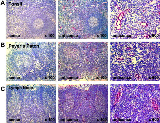
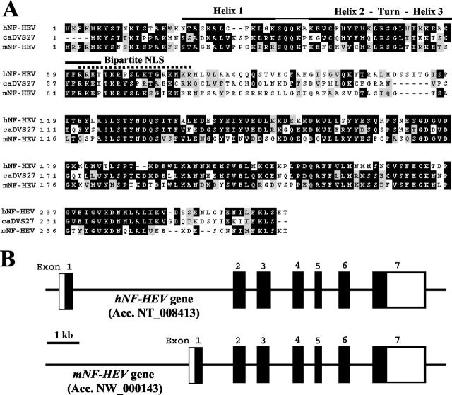
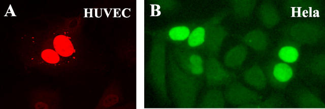
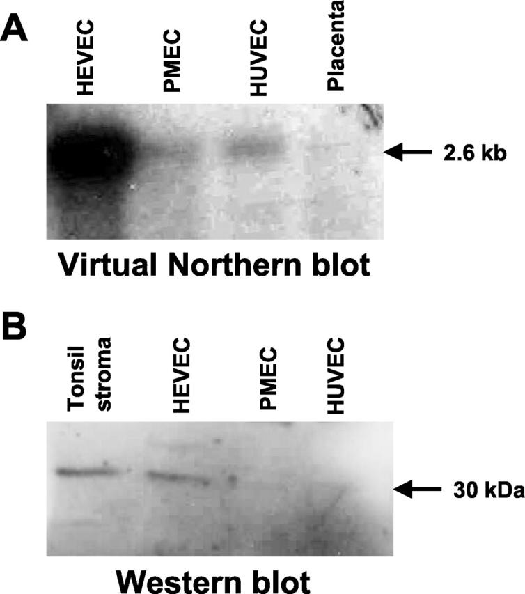
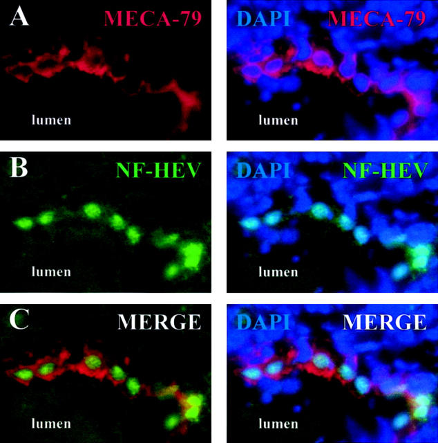
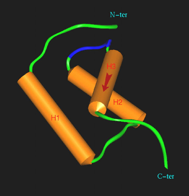
Lymphocyte homing to secondary lymphoid tissue and lesions of chronic inflammation is directed by multi-step interactions between the circulating cells and the specialized endothelium of high endothelial venules (HEVs). In this study, we used the PCR-based method of suppression subtractive hybridization (SSH) to identify novel HEV genes by comparing freshly purified HEV endothelial cells (HEVECs) with nasal polyp-derived microvascular endothelial cells (PMECs). By this approach, we cloned the first nuclear factor preferentially expressed in HEVECs, designated nuclear factor from HEVs (NF-HEV). Virtual Northern and Western blot analyses showed strong expression of NF-HEV in HEVECs, compared to human umbilical vein endothelial cells (HUVECs) and PMECs. In situ hybridization and immunohistochemistry revealed that NF-HEV mRNA and protein are expressed at high levels and rather selectively by HEVECs in human tonsils, Peyers's patches, and lymph nodes. The NF-HEV protein was found to contain a bipartite nuclear localization signal, and was targeted to the nucleus when ectopically expressed in HUVECs and HeLa cells. Furthermore, endogenous NF-HEV was found in situ to be confined to the nucleus of tonsillar HEVECs. Finally, threading and molecular modeling studies suggested that the amino-terminal part of NF-HEV (aa 1-60) corresponds to a novel homeodomain-like Helix-Turn-Helix (HTH) DNA-binding domain. Similarly to the atypical homeodomain transcription factor Prox-1, which plays a critical role in the induction of the lymphatic endothelium phenotype, NF-HEV may be one of the key nuclear factors that controls the specialized HEV phenotype.


Figures

Figure 1.
Figure 1.
NF-HEV mRNA expression in HEVs of human tonsil, Peyer’s patch, and mesenteric lymph node. In situ hybridization was performed on paraformaldehyde-fixed sections with an RNA probe complementary to NF-HEV mRNA (antisense), and hybridization signal (red) occurs in HEVs of the T-cell zone around lymphoid follicles in tonsil (A), Peyer’s patch (B), and mesenteric lymph node (C). Higher magnification (×600, right panels) reveals that the signal is confined to HEVECs and scattered cells in the T- and B-cell zones. Hybridization with a sense probe produced no signal (left panels).
Figure 2.
Figure 2.
Human and mouse NF-HEV proteins and genes. A: Amino acid sequence alignment of human NF-HEV (hNF-HEV) with its mouse ortholog (mNF-HEV) and canine DVS27 (caDVS27). Conserved residues are boxed. Black boxes indicate identical residues, whereas shaded boxes show similar amino acids. Dashed lines represent gaps introduced to align sequences. Sequence alignment was performed with ClustalW (http://www2.ebi.ac.uk/clustalw) and colored with Boxshade (http://www.ch.embnet.org/software/BOX_form.html). The bipartite NLS and the three helices of the homeodomain-like HTH putative DNA-binding motif are indicated. B: Genomic structure of the human and mouse NF-HEV genes. Open boxes indicate non-translated exon sequence and black boxes coding exon sequence. The two genes share a similar organization with seven exons. A major difference is the size of the first intron, which is >9 kb in the human gene but only ∼2 kb in its mouse ortholog.
Figure 3.
Figure 3.
NF-HEV encodes a nuclear protein. A–B: Nuclear localization of epitope-tagged NF-HEV ectopically expressed in primary HUVECs or immortalized HeLa epithelial cells. HUVECs (A) and HeLa cells (B) transfected with myc-tagged NF-HEV expression vector were stained by indirect immunofluorescence with antibodies to myc and analyzed by confocal laser scanning microscopy. Original magnification, ×1000.
Figure 4.
Figure 4.
Virtual Northern and Western blot analyses demonstrating preferential expression of NF-HEV in HEVECs. A: Virtual Northern blot analysis of NF-HEV expression in HEVECs, PMECs, HUVECs, or placenta tissue. PCR-generated full-length cDNAs from the various types of ECs were electrophoresed on a 1% agarose gel, transferred to nylon filters, and hybridized under high-stringency conditions with a 32P-labeled human NF-HEV cDNA probe. B: Western blot analysis of extracts of tonsillar stroma, HEVECs, PMECs, or HUVECs with rabbit antibodies to NF-HEV. A single band of ∼30 kd was detected in extracts of tonsillar stroma and HEVECs.
Figure 5.
Figure 5.
In situ expression of NF-HEV protein in the nucleus of tonsillar HEVECs. Cryosections of human tonsils (4 μm, acetone-fixed) were double-stained with HEV-specific rat mAb MECA-79 (A) or antibodies to NF-HEV peptides (B). C: Two-color overlays reveal that NF-HEV immunoreactivity is associated with MECA-79-positive HEVECs. Counterstaining with the nuclear dye DAPI showed a clear nuclear localization of NF-HEV in MECA-79-positive HEVECs (right panels). No nuclear staining was observed with preimmune rabbit serum (not shown). Original magnification, ×600.
Figure 6.
Figure 6.
NF-HEV contains a homeodomain-like HTH motif. Model of the three-dimensional structure of the homeodomain-like HTH motif of human NF-HEV (aa 1–65), based on its threading-derived homology with the crystallographic structure of the homeodomain from drosophila transcription factor engrailed (PDB code: 1DU0). The α-helices have been numbered in order and color-coded in brown. The potential DNA recognition helix (α-helix 3) is marked by a red arrow. The turn of the HTH motif is coded in blue. Molecular modeling was performed as described in the Materials and Methods.

References

    1. Cines DB, Pollak ES, Buck CA, Loscalzo J, Zimmerman GA, McEver RP, Pober JS, Wick TM, Konkle BA, Schwartz BS, Barnathan ES, McCrae KR, Hug BA, Schmidt AM, Stern DM: Endothelial cells in physiology and in the pathophysiology of vascular disorders. Blood 1998, 91:3527-3561 - PubMed
    1. Risau W: Differentiation of endothelium. EMBO J 1995, 9:926-933 - PubMed
    1. Girard JP, Springer TA: High endothelial venules (HEVs): specialized endothelium for lymphocyte migration. Immunol Today 1995, 16:449-457 - PubMed
    1. Kraal G, Mebius RE: High endothelial venules: lymphocyte traffic control and controlled traffic. Adv Immunol 1997, 65:347-395 - PubMed
    1. Freemont AJ: Molecules controlling lymphocyte-endothelial interactions in lymph nodes are produced in vessels of inflamed synovium. Ann Rheum Dis 1987, 46:924-928 - PMC - PubMed

Publication types

MeSH terms