. 2003 Jul;163(1):91-100.
doi: 10.1016/s0002-9440(10)63633-4.

Ubiquitination of alpha-synuclein is not required for formation of pathological inclusions in alpha-synucleinopathies

Affiliations

Ubiquitination of alpha-synuclein is not required for formation of pathological inclusions in alpha-synucleinopathies

Deepak M Sampathu et al. Am J Pathol. 2003 Jul.

Abstract

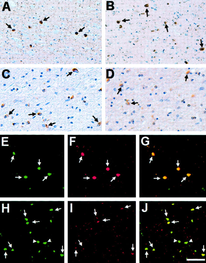
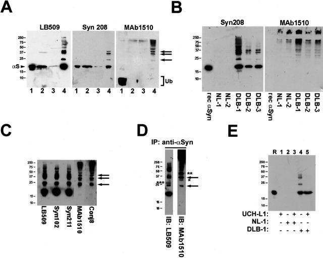
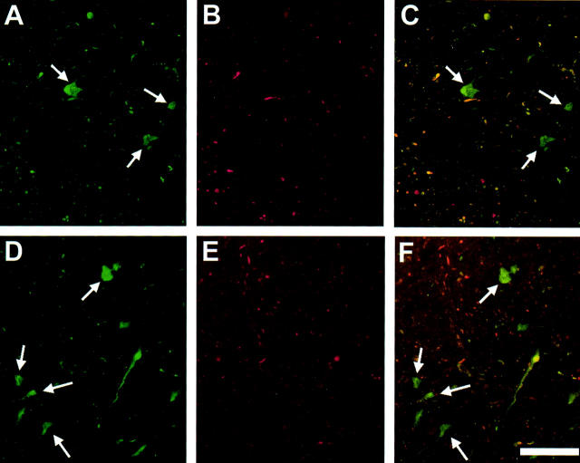
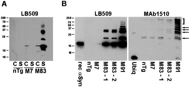
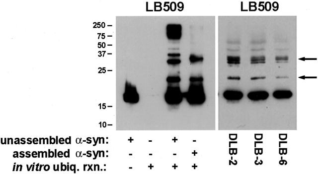
alpha-Synucleinopathies, including Parkinson's disease, dementia with Lewy bodies, and multiple system atrophy, are neurodegenerative disorders in which abnormal inclusions containing alpha-synuclein accumulate in selectively vulnerable neurons and glia. In this report, immunohistochemistry demonstrates ubiquitin in subsets of alpha-synuclein inclusions in dementia with Lewy bodies and multiple system atrophy. Biochemistry demonstrates that alpha-synuclein in the sodium dodecyl sulfate-soluble fractions of diseased brains is ubiquitinated, with mono- and di-ubiquitinated species predominating over polyubiquitinated forms. Similar immunohistochemical and biochemical characteristics were observed in an A53T mutant human alpha-synuclein transgenic mouse model of neurodegenerative alpha-synucleinopathies. Furthermore, in vitro ubiquitination of alpha-synuclein fibrils recapitulated the pattern of alpha-synuclein ubiquitination observed in human disease and the A53T alpha-synuclein mouse model. These results suggest that ubiquitination of alpha-synuclein is not required for inclusion formation and follows the fibrillization of alpha-synuclein.


Figures

Figure 1.
Figure 1.
Ubiquitin immunostaining of α-syn pathological inclusions in DLB and MSA. Immunohistochemistry of cingulate cortex from a DLB patient (DLB-2) (A and B) and cerebellum from a MSA patient (MSA-6) (C and D) stained using monoclonal anti-α-syn antibodies Syn303 (A and C) and monoclonal anti-ubiquitin antibody mAb 1510 (B and D). In A and B, arrows highlight immunoreactive cortical LBs, whereas in C and D arrows indicate stained GCIs. Double-label immunofluorescence of cortical LBs in the cingulate cortex of a patient with DLB (DLB-5) (E–G) and GCIs in the cerebellum of a patient with MSA (MSA-8) (H–J) with rabbit anti-α-syn antibody SNL-4 (green, E and H) and murine anti-ubiquitin antibody mAb 1510 (red, F and I). The overlays of staining with both antibodies are shown in G and J. In H to J, arrows indicate inclusions stained with antibodies to both α-syn and ubiquitin, whereas arrowheads depict inclusions stained only with antibodies to α-syn. Scale bars: 80 μm (A, B, E–J), 40 μm (C, D).
Figure 2.
Figure 2.
Insoluble α-syn in diseased brain is ubiquitinated. A: Western blot analysis of biochemically fractionated cingulate cortex from a patient with DLB (DLB-3). Immunoblots were developed with anti-α-syn antibodies LB509 and Syn208 as well as anti-ubiquitin antibody mAb 1510. Twenty μl of LS fraction (lane 1), TX fraction (lane 2), sarkosyl-soluble fraction (lane 3), and SDS-soluble fraction (lane 4) were loaded in separate lanes of 15% SDS-polyacrylamide gels. Note that the SDS-soluble fraction is four times as concentrated as each of the other fractions in that 2.5 ml/g of SDS buffer was used for tissue extraction versus 10 ml/g for each of the other fractions. Arrowhead indicates α-syn monomer (αS) and bracket indicates ubiquitin monomer (Ub). Arrows depict mono-, di-, and tri-ubiquitinated forms of α-syn. B: Western blot analysis (with antibodies Syn208 and mAb 1510) of the SDS-soluble fraction from the cingulate cortex of normal brains (NL-1, NL-2) and DLB brains (DLB-1, DLB-2, and DLB-3). Twenty μl of SDS-soluble fraction was loaded in each lane of a 15% gel. One hundred ng of recombinant human α-syn was loaded in the indicated lanes. C: Western blot analysis of the SDS-soluble fraction from the cingulate cortex of case DLB-1 using various anti-α-syn (LB509, Syn102, Syn211) and anti-ubiquitin (mAb 1510, Conj8) antibodies. Arrows depict mono-, di-, and tri-ubiquitinated forms of α-syn. D: Immunoprecipitation followed by Western blot analysis. α-Syn in the SDS-soluble fraction of the cingulate cortex of DLB-1 was isolated by immunoprecipitation with anti-α-syn antibodies. The sample was analyzed by Western blot analysis using anti-α-syn antibody LB509 and anti-ubiquitin antibody mAb 1510 (arrows, mono- and di-ubiquitinated forms of α-syn; * and **, possible ubiquitinated forms of α-syn in which ubiquitin moieties may be masking the LB509 epitope; ***, modified form of α-syn, possibly dimerized α-syn). E: Ubiquitinated α-syn from DLB brain can be deubiquitinated by UCH-L1 in vitro. SDS-soluble fraction from the cingulate cortex of NL-1 or DLB-1 was untreated or reacted with 50 nmol/L of UCH-L1. The samples were analyzed by Western blot analysis using LB509. R represents a lane loaded with 100 ng of recombinant human α-syn. The mobility of molecular mass markers (kd) is depicted on the left of each panel.
Figure 3.
Figure 3.
Ubiquitin immunostaining of α-syn inclusions in mice expressing A53T human α-syn. Double-label immunofluorescence of α-syn inclusions in the spinal cord (A–C) and pons (D–F) of M83 homozygous A53T human α-syn transgenic mice with rabbit anti-α-syn antibody SNL-4 (green, A and D) and mouse anti-ubiquitin antibody mAb 1510 (red, B and E). The overlays are shown in C and F. Arrows indicate α-syn inclusions that are not ubiquitin-positive. Scale bar, 80 μm.
Figure 4.
Figure 4.
α-Syn is ubiquitinated in inclusions in A53T α-syn transgenic mice. A: Western blot analysis of the SDS-soluble fractions of cortex (C) and spinal cord (S) from 12-month-old nontransgenic (nTg) mice, homozygous transgenic mice expressing wild-type human α-syn (line M7), and homozygous transgenic mice expressing A53T human α-syn (line M83) using the anti-α-syn antibody LB509. Note the accumulation of α-syn and higher molecular mass species in the SDS-soluble fraction of M83 transgenic mice. B: Accumulation of higher molecular mass species of α-synuclein in the spinal cord of M83 and M91 transgenic mice coincides with presence of ubiquitin immunoreactive bands. Ten μl of each SDS-soluble fraction was loaded in separate lanes of a 15% polyacrylamide gel. Arrows indicate ubiquitinated forms of α-syn in the M91 mouse. The bracket indicates ubiquitinated proteins that are unlikely to be α-syn because they are not labeled by the anti-α-syn antibody LB509. The mobility of molecular mass markers (kd) is indicated on the left of each panel.
Figure 5.
Figure 5.
In vitro ubiquitination of monomeric and fibrillar α-syn. Left: Unassembled or fibrillar α-syn (0.5 mg/ml) was subjected to in vitro ubiquitination as described in Materials and Methods. Equal volumes of unassembled α-syn, ubiquitination reaction without α-syn, ubiquitination reaction with unassembled α-syn, and ubiquitination reaction with fibrillar α-syn were loaded in separate lanes of 15% SDS-polyacrylamide gels that were transferred electrophoretically onto nitrocellulose and analyzed by Western blotting with LB509. Right: Western blot analysis (using LB509) of the SDS-soluble fraction of cingulate cortex from three DLB cases. Arrows indicate ubiquitinated forms of α-syn. The mobility of molecular mass markers (kd) is depicted to the left of the panel.

References

    1. Spillantini MG, Goedert M: The α-synucleinopathies: Parkinson’s disease, dementia with Lewy bodies, and multiple system atrophy. Ann NY Acad Sci 2000, 920:16-27 - PubMed
    1. Duda JE, Lee VM-Y, Trojanowski JQ: Neuropathology of synuclein aggregates. J Neurosci Res 2000, 61:121-127 - PubMed
    1. Goedert M: α-Synuclein and neurodegenerative diseases. Nat Rev Neurosci 2001, 2:492-501 - PubMed
    1. Polymeropoulos MH, Lavedan C, Leroy E, Ide SE, Dehejia A, Dutra A, Pike B, Root H, Rubenstein J, Boyer R, Stenroos ES, Chandrasekharappa S, Athanassiadou A, Papapetropoulos T, Johnson WG, Lazzarini AM, Duvoisin RC, Di Iorio G, Golbe LI, Nussbaum RL: Mutation in the α-synuclein gene identified in families with Parkinson’s disease. Science 1997, 276:2045-2047 - PubMed
    1. Kruger R, Kuhn W, Muller T, Woitalla D, Graeber M, Kosel S, Przuntek H, Epplen JT, Schols L, Riess O: Ala30Pro mutation in the gene encoding α-synuclein in Parkinson’s disease. Nat Genet 1998, 18:106-108 - PubMed

Publication types

MeSH terms