. 2003 Jul;163(1):121-33.
doi: 10.1016/s0002-9440(10)63636-x.

Mononuclear leukocytes bind to specific hyaluronan structures on colon mucosal smooth muscle cells treated with polyinosinic acid:polycytidylic acid: inter-alpha-trypsin inhibitor is crucial to structure and function

Affiliations

Mononuclear leukocytes bind to specific hyaluronan structures on colon mucosal smooth muscle cells treated with polyinosinic acid:polycytidylic acid: inter-alpha-trypsin inhibitor is crucial to structure and function

Carol A de la Motte et al. Am J Pathol. 2003 Jul.

Abstract

Inflammatory bowel disease (IBD) is a chronic disorder whose etiology is linked to triggering events, including viral infections, that lead to immunoregulatory dysfunction in genetically susceptible people. Characteristic pathological changes include increased mononuclear leukocyte influx into the intestinal mucosa as well as mucosal smooth muscle cell (M-SMC) hyperplasia. Virus infection or viral mimic [polyinosinic acid:polycytidylic acid (polyI:C)] treatment of human colon M-SMCs in vitro increases cell surface hyaluronan (HA), and nonactivated mononuclear leukocytes bind to virus-induced HA structures by interactions that involve the HA-binding receptor CD44. In this study, confocal microscopy reveals increased HA on poly I:C-treated M-SMC surfaces within 3 hours, arrayed in coat-like structures. By 17 hours, novel, lengthy cable structures are evident, and these are primarily responsible for mediating leukocyte adhesion. Immunohistochemical staining demonstrates components of the inter-alpha-trypsin inhibitor (IalphaI) complex in both coat-like and cable structures. M-SMCs co-treated with polyI:C and a polyclonal antibody to IalphaI display HA in coats but with diminished cables, and they bind significantly fewer leukocytes than M-SMCs treated with polyI:C alone. Western blot data suggest that heavy chains of IalphaI are specifically associated with cable structures. Staining of tissue sections from patients with IBD demonstrates the presence of HA in inflamed colon tissue, and shows that HA-associated IalphaI staining increases in the mucosa of inflamed IBD specimens compared to noninflamed sections from the same patient, establishing a probable link between the observations in vitro and the progression of the inflammatory process in IBD.


Figures

Figure 1.
Figure 1.
U937 cell binding to polyI:C-induced HA on M-SMCs occurs in clusters. Confluent M-SMCs were treated with DME/F12 medium containing 10% FBS with or without polyI:C for 18 hours at 37°C. U937 cell adhesion was done as described in Materials and Methods and observed by phase contrast microscopy (×100). U937 cells appear as bright spheres on top of the M-SMCs, which are attached to the culture plate. a, Medium treated; b, polyI:C treated; and c, polyI:C treated with hyaluronidase (200 μg/ml) added after adhesion followed by incubation at 25°C for 5 minutes and washing to remove the released U937 cells.
Figure 2.
Figure 2.
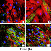
HA is displayed in two distinct structures on polyI:C-treated M-SMCs throughout time. Confluent M-SMCs grown on coverslips were treated with DME/F12 medium containing 10% FBS and polyI:C at 37°C for the times indicated. Coverslips were methanol fixed at intervals throughout 17 hours. Cells were then fluorescently labeled for detection of HA (green, secondary FITC-conjugated reagent), CD44 (red, secondary Texas Red-conjugated antibody), and nuclei (blue, DAPI) (described in Materials and Methods) and observed by confocal microscopy (×20 objective).
Figure 3.
Figure 3.
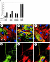
U937 cells bind to polyI:C-induced cable structures but not to coats on the surface of M-SMCs. Confluent M-SMCs grown on coverslips were treated with DME/F12 medium containing 10% FBS and polyI:C for 17 hours at 37°C. The U937 cell adhesion assay was done as described in Materials and Methods. The coverslips were then washed, methanol-fixed, and labeled for detection of HA (green, secondary FITC-conjugated reagent), CD44 (red, secondary Texas Red-conjugated antibody), and nuclei (blue, DAPI) (described in Materials and Methods) and observed by confocal microscopy. a: Many CD44-stained U937 cells are attached to the cables, which extend above the adherent monolayer of M-SMCs (×40 objective). Arrowheads indicate patches, which are devoid of leukocytes. b: A single U937 cell is bound to a cable (arrow) rising above the M-SMC nucleus, and not to the patches of HA associated with the M-SMC surface (×100 objective).
Figure 4.
Figure 4.
A: Effect of polyI:C on M-SMC expression of cell surface HA, IαI, versican, and CD44. Confluent M-SMCs were treated with DME/F12 medium containing 10% FBS with or without polyI:C for 18 hours at 37°C. Binding of the HA probe, or antibodies to IαI, versican, or CD44 was quantitated as described in Materials and Methods. Values are the mean of triplicate wells ± SEM. B: IαI is associated with HA on both the M-SMC surface and the polyI:C-induced cables. Confluent M-SMCs grown on coverslips were treated with DME/F12 medium with (a, b, d–f) or without (c) 10% FBS, and with (b–f) or without polyI:C (a) for 18 hours at 37°C and then methanol-fixed. Cells were then fluorescently labeled for detection of HA (green, secondary FITC-conjugated reagent), IαI (red, secondary Texas Red-conjugated antibody), and nuclei (blue, DAPI) (described in Materials and Methods) and observed by confocal microscopy (×63 objective). Yellow color indicates co-localization of red and green staining.
Figure 5.
Figure 5.
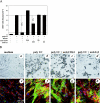
A: Effect of anti-CD44 and anti-IαI antibodies on polyI:C-induced U937 cell adhesion. Confluent M-SMCs were treated with DME/F12 medium containing 10% FBS with or without polyI:C (20 μg/ml). Replicate cultures were co-treated with polyI:C and CD44 antibody (20 μg/ml) or with polyI:C and IαI antibody (1:50). All cultures were incubated for 18 hours at 37°C. In some of the replicate polyI:C-treated cultures, CD44 antibody (10 μg/ml) or IαI antibody (1:50) were added 2 hours before the end of the incubation period. U937 cell adhesion was measured as described in Materials and Methods. A replicate set was also treated with hyaluronidase (200 μg/ml) added after adhesion followed by incubation at 25°C for 5 minutes and washing to remove the released U937 cells. Values are the mean of triplicate wells ± SEM. B: Effect of anti-CD44 and anti-IαI antibodies on polyI:C-induced HA cable formation. Confluent M-SMCs were treated with DME/F12 medium containing 10% FBS with or without polyI:C (20 μg/ml). Some replicate cultures were co-treated with polyI:C and CD44 antibody (10 μg/ml) or with polyI:C and IαI antibody (1:50). All cultures were incubated for 18 hours at 37°C. The U937 cell adhesion assay was done as described in Materials and Methods after which the coverslips were washed and methanol-fixed. Light microscopic images were obtained (a–d) and then cells were fluorescently labeled for detection of HA (green, secondary FITC-conjugated reagent), CD44 (red, secondary Texas Red-conjugated antibody), and nuclei (blue, DAPI) as described in Materials and Methods and observed by confocal microscopy (e–h) (×10 objective).
Figure 6.
Figure 6.
Western blot analysis for HA-associated IαI components in the cell layer of unstimulated and polyI:C-treated M-SMCs. Confluent M-SMCs were treated with DME/F12 medium containing 10% FBS with or without polyI:C (20 μg/ml) for 18 hours. Cells were rinsed and treated with Streptomyces hyaluronidase (100 mU/ml) for 5 minutes, processed, and Western blot analysis of IαI done as described in Materials and Methods. A: Whole M-SMC extracts from medium-treated (M), poly I:C-treated (P), or poly I:C-treated cells that received a limited hyaluronidase digestion (P + HAase), as well as the cell surface digest (HAase digest) were compared. B: Hyaluronidase digests from medium-treated (M) or polyI:C-treated (P) M-SMC cultures from three separate patient isolates, hyaluronidase solution alone (H) or containing fetal bovine serum (S) were compared to two different molecular weight standards (Std #1 and Std #2). C: Hyaluronidase (HA’ase) digests from medium-treated (M) or polyI:C-treated (P) M-SMC from a single patient were treated with chondroitinase ABC (C’ase) or NaOH as described in Materials and Methods and processed for Western blot analysis.
Figure 7.
Figure 7.
Detection of HA and CD44 in tissue sections from noninflamed and inflamed human colon. Paraffin sections obtained from normal-appearing (a) or inflamed (b) segments of a resected colon from a patient with ulcerative colitis were fluorescently labeled for detection of HA (green, secondary FITC-conjugated reagent) and nuclei (blue, DAPI) (described in Materials and Methods) and observed by confocal microscopy. Paraffin sections obtained from the same inflamed ulcerative colitis tissue or mildly inflamed (d) or inflamed (e) segments of a resected colon from a different patient with Crohn’s disease were fluorescently labeled for detection of HA (green, secondary FITC-conjugated reagent), CD44 (red, secondary Texas Red-conjugated antibody), and nuclei (blue, DAPI) (described in Materials and Methods) and observed by confocal microscopy [×10 objective (a, b, d); ×40 objective (c, e)].
Figure 8.
Figure 8.
Detection of IαI and HA in tissue sections from noninflamed and inflamed human colon. Paraffin sections obtained from normal appearing (a) or inflamed (b) segments of a resected colon from a patient with ulcerative colitis were fluorescently labeled for detection of HA (green, secondary FITC-conjugated reagent), IαI (red, secondary Texas Red-conjugated antibody), and nuclei (blue, DAPI) (described in Materials and Methods) and observed by confocal microscopy (×10 objective). Higher magnification (×63 objective–zoom 1) of the muscularis mucosae layer in b shows individual detail of green HA staining (c), red IαI staining (d), and the overlay of the two (e) plus blue DAPI nuclear stain to help determine the location of the individual M-SMCs. Yellow color indicates co-localization of red and green staining.

References

    1. Kangro HO, Chong SK, Hardiman A, Heath RB, Walker-Smith JA: A prospective study of virus and mycoplasm infections in chronic inflammatory bowel disease. Gastroenterology 1990, 98:549-553 - PubMed
    1. Yanai H, Shimizu N, Nagasaki S, Mitani N, Okita K: Epstein-Barr virus infection of the colon with inflammatory bowel disease. Am J Gastroenterology 1999, 98:549-553 - PubMed
    1. de la Motte CA, Hascall VC, Calabro A, Yen-Lieberman B, Strong SA: Mononuclear leukocytes preferentially bind via CD44 to hyaluronan on human intestinal mucosal smooth muscle cells after virus infection or treatment with poly I: C. J Biol Chem 1999, 274:30747-30755 - PubMed
    1. Aruffo A, Stamenkovic I, Melnick M, Underhill CB, Seed B: CD44 is the principal cell surface receptor for hyaluronate. Cell 1990, 61:1303-1313 - PubMed
    1. Jain M, He Q, Lee W-S, Kashiki S, Foster LC, Tsai J-C, Lee M-E, Haber E: Role of CD44 in the reaction of vascular smooth muscle cells to arterial wall injury. J Clin Invest 1996, 97:596-603 - PMC - PubMed

Publication types

MeSH terms