

The clinical significance of cathepsin S expression in human astrocytomas
- PMID: 12819022
- PMCID: PMC1868175
- DOI: 10.1016/S0002-9440(10)63641-3
The clinical significance of cathepsin S expression in human astrocytomas
Abstract
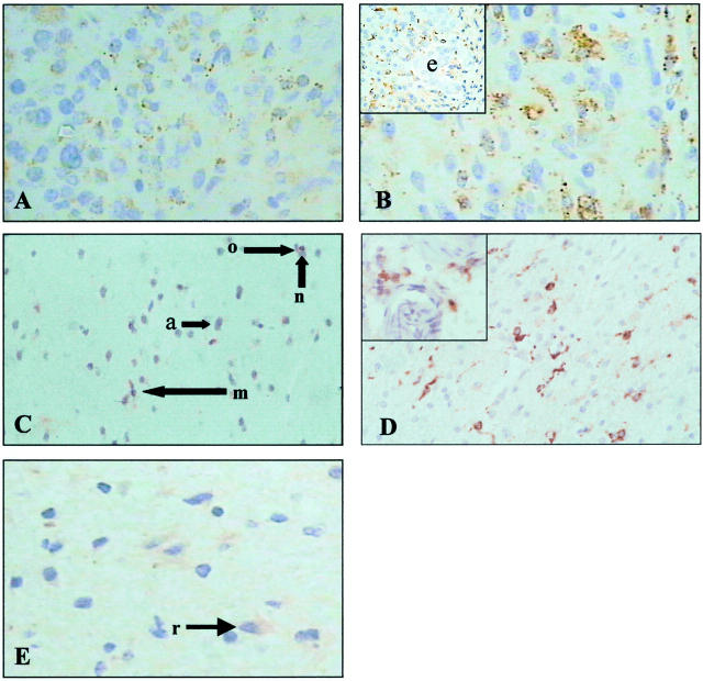
Early local invasion by astrocytoma cells results in tumor recurrence even after apparent total surgical resection, leading to the poor prognosis associated with malignant astrocytomas. Proteolytic enzymes have been implicated in facilitating tumor cell invasion and the current study was designed to characterize the expression of the cysteine proteinase cathepsin S (CatS) in astrocytomas and examine its potential role in invasion. Immunohistochemical analysis of biopsies demonstrated that CatS was expressed in astrocytoma cells but absent from normal astrocytes, oligodendrocytes, neurones and endothelial cells. Microglial cells and macrophages were also positive. Assays of specific activity in 59 astrocytoma biopsies confirmed CatS expression and in addition demonstrated that the highest levels of activity were expressed in grade IV tumors. CatS activity was also present in astrocytoma cells in vitro and the extracellular levels of activity were highest in cultures derived from grade IV tumors. In vitro invasion assays were carried out using the U251MG cell line and the invasion rate was reduced by up to 61% in the presence of the selective CatS inhibitor 4-Morpholineurea-Leu-HomoPhe-vinylsulphone. We conclude that CatS expression is up-regulated in astrocytoma cells and provide evidence for a potential role for CatS in invasion.
Figures


Similar articles
-
Detection of cathepsin S cysteine protease in human brain tumour microdialysates in vivo.Br J Neurosurg. 2007 Apr;21(2):204-9. doi: 10.1080/02688690701248190. Br J Neurosurg. 2007. PMID: 17453790 Clinical Trial.
-
Cathepsin S expression: An independent prognostic factor in glioblastoma tumours--A pilot study.Int J Cancer. 2006 Aug 15;119(4):854-60. doi: 10.1002/ijc.21911. Int J Cancer. 2006. PMID: 16550604
-
Lysosomal enzymes, cathepsins in brain tumour invasion.J Neurooncol. 2002 May;58(1):21-32. doi: 10.1023/a:1015892911420. J Neurooncol. 2002. PMID: 12160137 Review.
-
Expression pattern of alpha-protein kinase C in human astrocytomas indicates a role in malignant progression.Cancer Res. 1992 May 15;52(10):2951-6. Cancer Res. 1992. PMID: 1316231
-
Pathologic analysis of primary brain tumors.Neurol Clin. 1985 Nov;3(4):711-28. Neurol Clin. 1985. PMID: 3001488 Review.
Cited by
-
Evaluating the diagnostic and prognostic value of circulating cathepsin S in gastric cancer.Oncotarget. 2016 May 10;7(19):28124-38. doi: 10.18632/oncotarget.8582. Oncotarget. 2016. PMID: 27058412 Free PMC article.
-
Cathepsin S attenuates endosomal EGFR signalling: A mechanical rationale for the combination of cathepsin S and EGFR tyrosine kinase inhibitors.Sci Rep. 2016 Jul 8;6:29256. doi: 10.1038/srep29256. Sci Rep. 2016. PMID: 27387133 Free PMC article.
-
Cathepsin B, D and S as Potential Biomarkers of Brain Glioma Malignancy.J Clin Med. 2022 Nov 15;11(22):6763. doi: 10.3390/jcm11226763. J Clin Med. 2022. PMID: 36431239 Free PMC article.
-
The role of Cathepsin S as a marker of prognosis and predictor of chemotherapy benefit in adjuvant CRC: a pilot study.Br J Cancer. 2011 Nov 8;105(10):1487-94. doi: 10.1038/bjc.2011.408. Epub 2011 Oct 11. Br J Cancer. 2011. PMID: 21989182 Free PMC article. Clinical Trial.
-
Protease-activated receptors in health and disease.Physiol Rev. 2023 Jan 1;103(1):717-785. doi: 10.1152/physrev.00044.2021. Epub 2022 Jul 28. Physiol Rev. 2023. PMID: 35901239 Free PMC article. Review.
References
-
- Daumas-Duport C, Scheithauer BW, Kelly T: A histologic and cytologic method for the spatial definition of gliomas. Mayo Clin Proc 1987, 62:435-439 - PubMed
-
- Silbergeld DL, Chicoine MR: Isolation and characterization of human malignant glioma cells from histologically normal brain. J Neurosurg 1997, 86:525-531 - PubMed
-
- Liotta LA: Tumor invasion and metastases role of the extracellular-matrix: Rhoads memorial award lecture. Cancer Res 1986, 46:1-7 - PubMed
-
- Rooprai HK, McCormick D: Proteases and their inhibitors in human brain tumours: a review. Anticancer Res 1997, 17:4151-4162 - PubMed
-
- McCormick D: Secretion of Cathepsin B by human gliomas in vitro. Neuropathol Appl Neurobiol 1993, 19:146-151 - PubMed
Publication types
MeSH terms
Substances
LinkOut - more resources
Full Text Sources
Other Literature Sources
Medical
Miscellaneous
