. 2003 Jul;163(1):231-42.
doi: 10.1016/S0002-9440(10)63646-2.

Carbon monoxide induces cytoprotection in rat orthotopic lung transplantation via anti-inflammatory and anti-apoptotic effects

Affiliations

Carbon monoxide induces cytoprotection in rat orthotopic lung transplantation via anti-inflammatory and anti-apoptotic effects

Ruiping Song et al. Am J Pathol. 2003 Jul.

Abstract

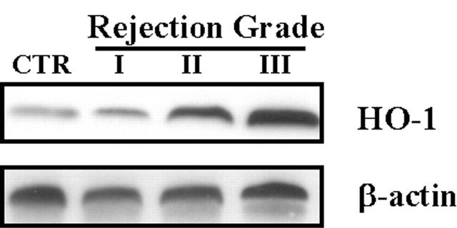
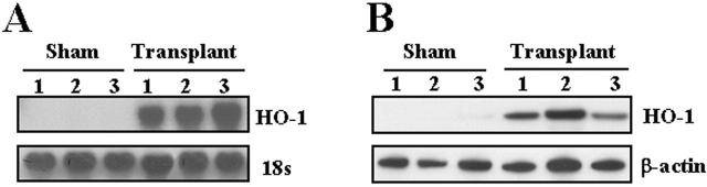
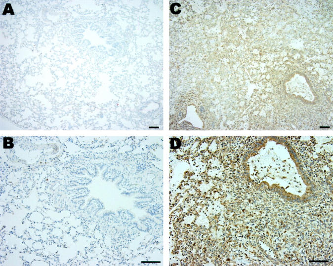
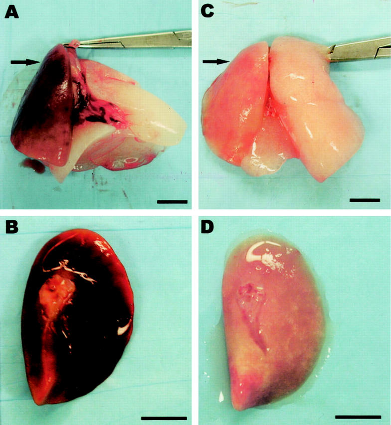
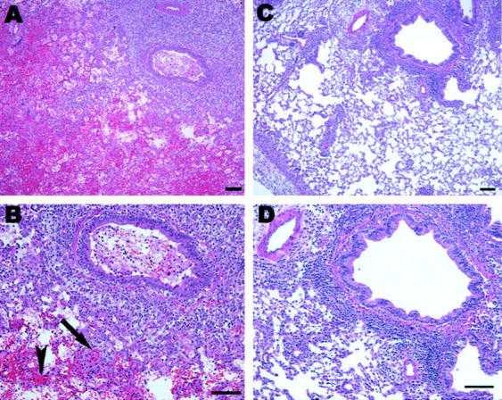
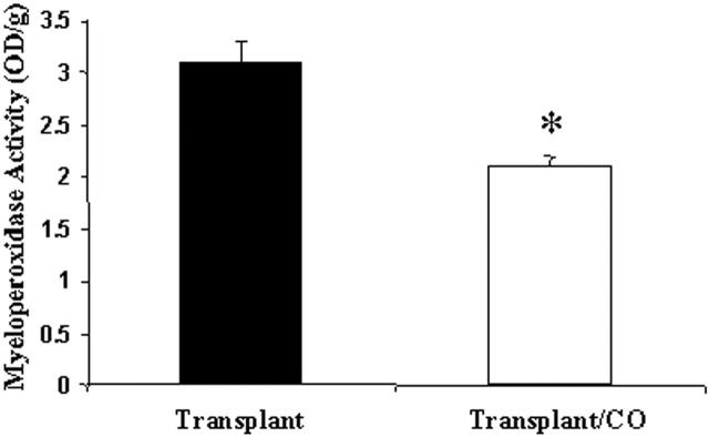
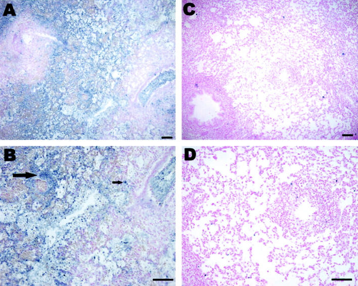
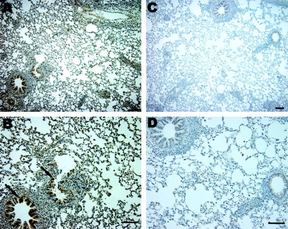
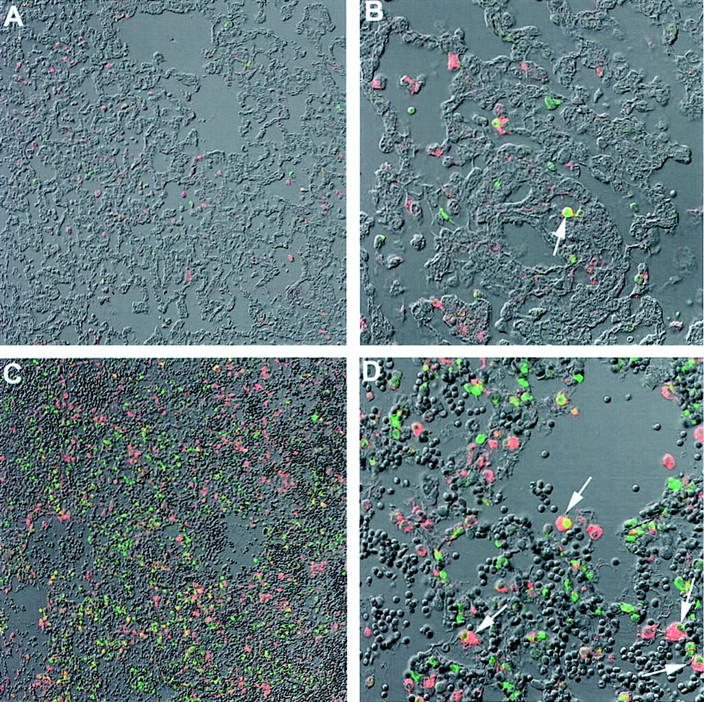
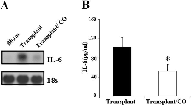
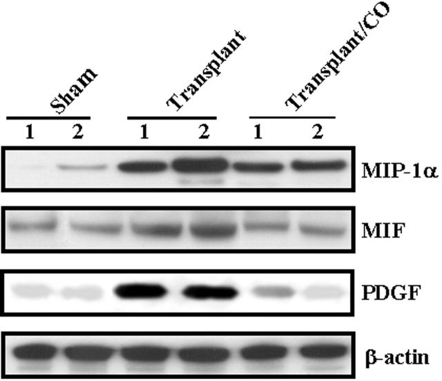
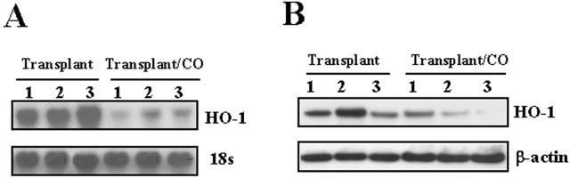
Successful lung transplantation has been limited by the high incidence of acute graft rejection. There is mounting evidence that the stress response gene heme oxygenase-1 (HO-1) and/or its catalytic by-product carbon monoxide (CO) confers cytoprotection against tissue and cellular injury. This led us to hypothesize that CO may protect against lung transplant rejection via its anti-inflammatory and antiapoptotic effects. Orthotopic left lung transplantation was performed in Lewis rat recipients from Brown-Norway rat donors. HO-1 mRNA and protein expression were markedly induced in transplanted rat lungs compared to sham-operated control lungs. Transplanted lungs developed severe intraalveolar hemorrhage, marked infiltration of inflammatory cells, and intravascular coagulation. However, in the presence of CO exposure (500 ppm), the gross anatomy and histology of transplanted lungs showed marked preservation. Furthermore, transplanted lungs displayed increased apoptotic cell death compared with the transplanted lungs of CO-exposed recipients, as assessed by TUNEL and caspase-3 immunostaining. CO exposure inhibited the induction of IL-6 mRNA and protein expression in lung and serum, respectively. Gene array analysis revealed that CO also down-regulated other proinflammatory genes, including MIP-1alpha and MIF, and growth factors such as platelet-derived growth factor, which were up-regulated by transplantation. These data suggest that the anti-inflammatory and antiapoptotic properties of CO confer potent cytoprotection in a rat model of lung transplantation.


Figures

Figure 1.
Figure 1.
HO-1 protein level in lung fibroblasts from human transplant recipients increased and correlated with acute rejection grade. Fibroblasts cultured from lung biopsy specimens of transplanted patients were harvested and cultured. Biopsy specimens were sent to pathology for acute rejection grading. Protein was extracted from cultured fibroblast and subjected to Western blot hybridization with HO-1 antibody as described in Methods. Each lane represents pooled samples from a group of five patients with acute rejection grade I to III; control was from normal patient lung. Cultured fibroblast was used at passage 2 to 3. The same membranes were probed with an antibody against β-actin to assure equal loading of the gel.
Figure 2.
Figure 2.
Rat lung transplantation increased HO-1 mRNA expression and protein level. A: Total RNA from the transplanted lung (day 4) was extracted and subjected to Northern blot hybridization with a 32P-labeled HO-1 cDNA probe as described in Methods. Each lane represents RNA extracted from one rat (n = 3). Normalization for RNA loading is shown by labeling 18S rRNA of the same membrane. B: Protein from the transplanted lung (day 4) was extracted and subjected to Western blot hybridization with HO-1 antibody as described in Methods. Each lane represents protein extracted from one rat (n = 3). The same membranes were probed with an antibody against β-actin to assure equal loading of the gel.
Figure 3.
Figure 3.
Expression of HO-1 in transplantation. Immunohistochemical staining for HO-1 demonstrated markedly elevated HO-1 expression (brown) in the transplanted lung section (C and D) compared to sham-operated lung section (A and B). Bar equals 100 μm.
Figure 4.
Figure 4.
Gross anatomy of lungs from rats 6 days after transplantation (arrow in A, B), and lungs from rats 6 days after transplantation which received 500 ppm CO over this time period (arrow in C, D). In A and C, the arrow points to the left transplanted lung; the right lung is the remaining native lung. Note that in the absence of CO, transplanted lungs were noted to be markedly hyperemic (A and B), which was notably absent in the transplanted lung exposed to CO (C and D). Scale bar, 1 cm.
Figure 5.
Figure 5.
H&E-stained sections of lungs from rats 6 days after transplantation (A and B) and lungs from rats 6 days after transplantation that received 500 ppm CO over this time period (C and D). Note: rejection is present in both CO-treated and untreated transplanted lungs as made evident by the pronounced perivascular and peribronchiole lymphocyte aggregates. However, lung transplant in the absence of CO were noted to have severe intraalveolar hemorrhage (arrowhead in B) and intravascular coagulation (arrow in B) that were notably absent in the transplanted lung exposed to CO (500 ppm) (C and D). Scale bar, 100 μm.
Figure 6.
Figure 6.
Myeloperoxidase activity was decreased in the CO-treated lung from transplantation. Rat lungs (n = 3) from both transplant and transplant/CO group were measured for MPO activity as described in Methods. MPO activity is expressed per gram of dry weight. CO treatment decreased MPO level by 31%. (* P < 0.05 versus control).
Figure 7.
Figure 7.
Effect of CO on transplantation-induced TUNEL staining. Markedly elevated TUNEL staining (A, arrow and blue staining in B) occurred in lungs from rats 6 days after transplantation compared to lungs (C and D) from transplantation which received 500 ppm CO over an equivalent time period. Scale bar, 100 μm.
Figure 8.
Figure 8.
Effect of CO on transplantation-induced activated caspase-3 expression. Immunohistochemical staining demonstrated markedly elevated activated caspase-3 expression (A, arrow and brown staining in B) in lungs from rats 4 days after transplantation compared to lungs (C and D) from transplantation which received 500 ppm CO over an equivalent time period. Scale bar, 100 μm.
Figure 9.
Figure 9.
TUNEL/macrophage co-staining. Sections were co-stained for TUNEL (green) and macrophage (red). A and B: Lung from rats 6 days after transplantation in the presence of CO (500 ppm). C and D: Lung from transplantation in the absence of CO. Arrows in B and D: Double-positive staining. We demonstrated markedly elevated co-staining for TUNEL/macrophage staining in lungs from rats after transplantation (C and D) compared to lungs from transplantation that received CO (500 ppm; A and B). Magnifications: A and C, ×20; B and D, ×60.
Figure 10.
Figure 10.
CO inhibited transplantation-induced IL-6 mRNA expression and serum IL-6. A: Total RNA from the transplanted lung was extracted and subjected to Northern blot hybridization with a 32P-labeled IL-6 cDNA probe as described in Methods. Each lane represents pooled RNA extracted from 3 rats (n = 3). Normalization for RNA loading is shown by labeling 18S rRNA of the same membrane. B: Serum was collected 4 days after transplantation and analyzed for IL-6 levels by ELISA. CO treatment decreased IL-6 level by 51%. (* P < 0.05 versus control). Data represent the means ± SE of samples from three independent experiments.
Figure 11.
Figure 11.
CO decreased protein levels of multiple chemokines/cytokines following lung transplantation. Western blots were performed to confirm changes in MIP-1α, MIF, and PDGF at the protein level, all of which demonstrated increase of mRNA expression in transplant group and decrease in transplant/CO group by cDNA array. Protein from the transplanted lung was extracted and subjected to Western blot hybridization with MIP-1α, MIF, and PDGF antibodies as described in Methods. The same membranes were probed with an antibody against β-actin to assure equal loading of the gel.
Figure 12.
Figure 12.
CO decreased HO-1 mRNA expression and protein level. A: Total RNA from the transplanted lung (day 4) was extracted and subjected to Northern blot hybridization with a 32P-labeled HO-1 cDNA probe as described in Methods. Each lane represents RNA extracted from 1 rat (n = 3). Normalization for RNA loading is shown by labeling 18S rRNA of the same membrane. B: Protein from the transplanted lung (day 4) was extracted and subjected to Western blot hybridization with HO-1 antibody as described in Methods. Each lane represents protein extracted from 1 rat (n = 3). The same membranes were probed with an antibody against β-actin to assure equal loading of the gel.

References

    1. Girgis RE, Tu I, Berry GJ, Reichenspurner H, Valentine VG, Conte JV, Ting A, Johnstone I, Miller J, Robbins RC, Reitz BA, Theodore J: Risk factors for the development of obliterative bronchiolitis after lung transplantation. J Heart Lung Transplant 1996, 15:1200-1208 - PubMed
    1. Bando K, Paradis IL, Similo S, Konishi H, Komatsu K, Zullo TG, Yousem SA, Close JM, Zeevi A, Duquesnoy RJ, Manzetti J, Keenan RJ, Armitage JM, Hardesty RL, Griffith BP: Obliterative bronchiolitis after lung and heart-lung transplantation: an analysis of risk factors and management. J Thorac Cardiovasc Surg 1995, 110:4-13 - PubMed
    1. Alwayn IP, Xu R, Adler WH, Kittur DS: Does high MHC class II gene expression in normal lungs account for the strong immunogenicity of lung allografts? Transpl Int 1994, 7:43-46 - PubMed
    1. Sibley RK, Berry GJ, Tazelaar HD, Kraemer MR, Theodore J, Marshall SE, Billingham ME, Starnes VA: The role of transbronchial biopsies in the management of lung transplant recipients. J Heart Lung Transplant 1993, 12:308-324 - PubMed
    1. Trulock EP: Management of lung transplant rejection. Chest 1993, 103:1566-1576 - PubMed

Publication types

MeSH terms