. 2003 Jul;163(1):303-12.
doi: 10.1016/S0002-9440(10)63654-1.

Blockade of the EGF receptor induces a deranged chemokine expression in keratinocytes leading to enhanced skin inflammation

Affiliations

Blockade of the EGF receptor induces a deranged chemokine expression in keratinocytes leading to enhanced skin inflammation

Francesca Mascia et al. Am J Pathol. 2003 Jul.

Abstract

During inflammatory skin disorders such as psoriasis, atopic dermatitis, and allergic contact dermatitis, epidermal keratinocytes overexpress large amounts of soluble epidermal growth factor receptor ligands in response to tumor necrosis factor alpha and interferon gamma. These cytokines also promote de novo synthesis of numerous chemokines, including CCL2/MCP-1, CCL5/RANTES, CXCL10/IP-10, and CXCL8/IL-8, in turn responsible for the recruitment of different leukocyte populations. This study demonstrates that stimulation of EGFR down-regulates CCL2, CCL5, and CXCL10, while it increases CXCL8 expression in keratinocytes. Conversely, EGFR signaling blockade produces opposite effects, with increased CCL2, CCL5, and CXCL10, and reduced CXCL8 expression. In a mouse model of contact hypersensitivity, a single topical administration of a selective EGFR kinase blocker before antigen challenge results in a markedly enhanced immune response with increased chemokine expression and heavier inflammatory cell infiltrate. Targeting EGFR on epithelial cells may thus have profound impact on inflammatory and immune responses.


Figures

Figure 1.
Figure 1.
EGFR and TGF-α expression in normal and chronically inflamed skin. Expression of EGFR in the epidermis of a healthy donor (A). EGFR expression in lesional skin of chronic plaque psoriasis (B), chronic atopic dermatitis (C), and chronic allergic contact dermatitis (D). Very faint TGF-α immunoreactivity in the epidermis of a healthy donor (E). Strong TGF-α expression in the epidermis and dermis of lesional skin of plaque psoriasis (F), atopic dermatitis (G), and allergic contact dermatitis (H). Representative immunohistochemistry results from four healthy donors, five patients with psoriasis, five patients with atopic dermatitis, and three patients with allergic contact dermatitis. Original magnification, ×100.
Figure 2.
Figure 2.
TNF-α and IFN-γ induce metalloproteinase-mediated shedding of membrane TGF-α. Flow cytometric analysis of cell-associated pro-TGF-α following 15 minutes of stimulation with 100 ng/ml TNF-α or 100 units/ml IFN-γ (A). TGF-α shedding was prevented by pre-incubation with the broad metalloproteinase inhibitor Ilomastat (B). Soluble TGF-α was measured in culture supernatants by ELISA (C). Keratinocytes were treated with cytokines or medium alone (no treatment, n.t.) directly (white columns), or after 30 minutes pre-incubation with Ilomastat (black columns).
Figure 3.
Figure 3.
TNF-α and IFN-γ induce EGFR transactivation. EGFR tyrosine phosphorylation (PY) in keratinocytes treated for 30 minutes with medium (n.t.), 50 ng/ml TGF-α, 100 ng/ml TNF-α, or 100 units/ml IFN-γ directly (-), or following 30 minutes pre-incubation with PD168393, AG1296, anti-TGF-α neutralizing Ab, or Ilomastat (A). EGFR was immunoprecipitated (IP) using anti-EGFR Ab, and phospho-EGFR was analyzed by Western blot (WB) using anti-phospho-tyrosine (PY) Ab. PD168393 (PD16) abrogated cytokine-induced EGFR and ERK phosphorylation (P-ERK1/2), but did not affect IFN-γR1 phosphorylation (P-IFN-γR1) following stimulation with IFN-γ, or TRAF2 recruitment by TNFR1 in TNF-α-stimulated keratinocytes (B). The number on the right side of each panel represents the weight in kd of the immunodetected molecule(s). Representative results from five independent experiments.
Figure 4.
Figure 4.
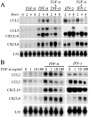
TGF-α down-regulates CCL2, CCL5, and CXCL10, but enhances CXCL8 expression. Time course (A) and dose dependence (B) of TGF-α effects on chemokine gene expression, as assessed by RNase protection assay. For the time course, TGF-α was used at a concentration of 50 ng/ml. The autoradiographies are representative of four independent experiments.
Figure 5.
Figure 5.
ELISA detection of chemokine levels after a 24-hour stimulation with escalating doses of TGF-α alone (white symbols), and together with TNF-α (▴) or IFN-γ (•) (C). Values represent ng/106 cells (± SD) from six independent experiments.
Figure 6.
Figure 6.
Impairment of EGFR signaling affects cytokine-induced chemokine expression with opposing effects compared to TGF-α, as observed by RNase protection assay. Cell cultures were pre-incubated with culture medium, PD168393, AG1296, neutralizing TGF-α Ab, or Ilomastat for 30 minutes, followed by stimulation with TNF-α for 2 hours (left panels) or IFN-γ for 8 hours (right panels). The autoradiographies are representative of four independent assays.
Figure 7.
Figure 7.
Chemokine release in the supernatants (24 hours) as assessed by ELISA. Values represent ng/106 cells (± SD) from six independent experiments. Cell cultures were pre-incubated with PD168393 or AG1296 for 30 minutes, followed by a further 24-hour treatment with TNF-α or IFN-γ, in the presence or not of TGF-α.
Figure 8.
Figure 8.
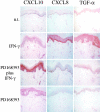
Immunohistochemistry of CXCL10, CXCL8, and TGF-α in cultures of full-thickness human skin explants. Skin explants were pre-incubated with vehicle alone (n.t.) or 2 μmol/L PD168393, and then stimulated or not with 1000 units/ml IFN-γ. Afterward, skin chops were dried by delicate contact with Whatman paper and snap-frozen. Representative results from three independent experiments. Original magnification, ×100.
Figure 9.
Figure 9.
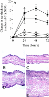
Treatment with PD168393 enhances contact hypersensitivity response. Contact hypersensitivity to DNFB was elicited on the ear skin of immunized BALB/c mice 30 minutes after local painting of 4 mmol/L PD168393 (▪) or vehicle alone (10% DMSO in absolute ethanol) (•). DNFB challenge solution was also applied to non-sensitized mice pre-treated with PD168393 (□) or vehicle alone (○) (A). Data represent the mean of the mean changes in ear thickness at each data point from five different experiments. *, P < 0.02 vs. the respective vehicle-treated group. Histological features of ear skin samples collected 48 hours after painting unsensitized mice with vehicle alone (B) or PD168393 (C), or 48 hours after DNFB challenge of sensitized mice pre-treated with vehicle alone (D) or PD168393 (E). Ear sections were stained with hematoxylin & eosin. Magnification, ×100.
Figure 10.
Figure 10.
In sensitized mice, a single topical administration of PD168393 30 minutes before challenge enhances keratinocyte-associated expression of CXCL10, CCL2, and CCL5, whereas it reduces TGF-α and phospho-ERK1/2, as assessed by immunohistochemical analyses (48 hours after challenge) (A). In control animals (n.t.), the site of DNFB challenge was pre-treated with vehicle alone. Original magnification, ×100. Total number of CD8+, CD4+ and CD11b+ leukocytes at the site of contact hypersensitivity 48 hours after treatment with vehicle alone (white columns) or PD168393 (black columns) and challenge with DNFB (B). Results represent the mean numbers (± SD) per mm2 (n = 6 microscopic fields per section, and are representative of three independent experiments. *, P < 0.02 vs. vehicle-treated group (n.t.).

References

    1. Sunnarborg SW, Hinkle CL, Stevenson M, Russell WE, Raska CS, Peschon JJ, Castner BJ, Gerhart MJ, Paxton RJ, Black RA, Lee DC: Tumor necrosis factor-α converting enzyme (TACE) regulates epidermal growth factor receptor ligand availability. J Biol Chem 2002, 277:12838-12845 - PubMed
    1. Stoll SW, Kansra S, Peshick S, Fry DW, Leopold WR, Wiesen JF, Sibilia M, Zhang T, Werb Z, Derinck R, Wagner EF, Elder JT: Differential utilization and localization of ErbB receptor tyrosine kinases in skin compared to normal and malignant keratinocytes. Neoplasia 2001, 3:339-350 - PMC - PubMed
    1. Schlessinger J: Ligand-induced, receptor-mediated dimerization and activation of EGF receptor. Cell 2002, 110:669-672 - PubMed
    1. Peus D, Hamacher L, Pittelkow MR: EGF-receptor tyrosine kinase inhibition induces keratinocyte growth arrest and terminal differentiation. J Invest Dermatol 1997, 109:751-756 - PubMed
    1. Hansen LA, Woodson RL, Holbus S, Strain K, Lo YC, Yuspa SH: The epidermal growth factor receptor is required to maintain the proliferative population in the basal compartment of epidermal tumors. Cancer Res 2000, 60:3328-3332 - PubMed

Publication types

MeSH terms