

Angiotensin II induces connective tissue growth factor gene expression via calcineurin-dependent pathways
- PMID: 12819040
- PMCID: PMC1868168
- DOI: 10.1016/S0002-9440(10)63659-0
Angiotensin II induces connective tissue growth factor gene expression via calcineurin-dependent pathways
Abstract
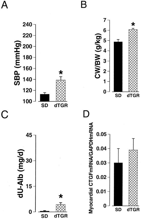
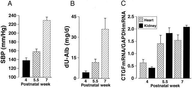
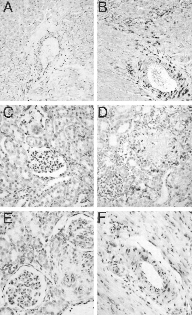
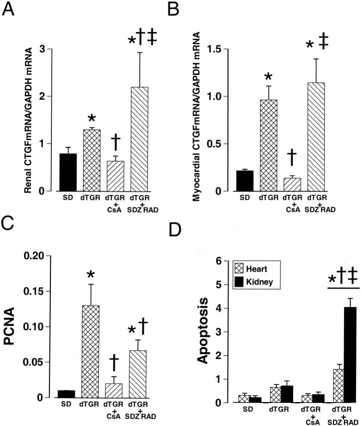
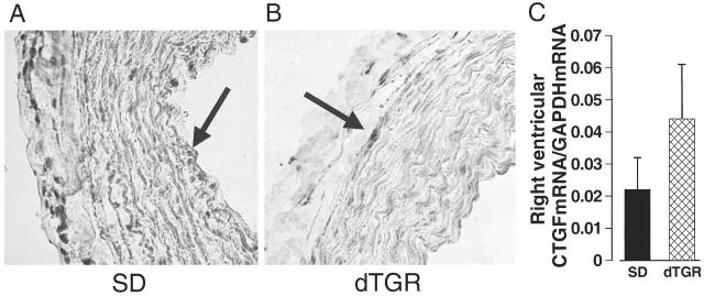
Connective tissue growth factor (CTGF) is a polypeptide implicated in the extracellular matrix synthesis. Previous studies have provided evidence that angiotensin II (Ang II) promotes collagen synthesis and regulates collagen degradation. We investigated whether or not CTGF mediates the profibrotic effects of Ang II in the heart and kidneys and the role of calcineurin-dependent pathways in CTGF gene regulation. In transgenic rats harboring human renin and angiotensinogen genes, Ang II induced an age-dependent increase in myocardial CTGF expression, which was 3.5-fold greater compared to normotensive Sprague Dawley (SD) rats. CTGF overexpression correlated closely with the Ang II-induced rise in blood pressure. CTGF mRNA and protein were located predominantly in areas with leukocyte infiltration, myocardial, and vascular lesions and co-localized with TGFbeta(1), collagen I, and collagen III mRNA expressions. Ang II induced CTGF mRNA and protein to a lesser extent in the kidneys, predominantly in glomeruli, arterioles, and in the interstitium with ample inflammation. However, no expression was found in the right ventricle or pulmonary arteries. Blockade of calcineurin activity by cyclosporine A completely normalized Ang II-induced CTGF overexpression in heart and kidney, suppressed the inflammatory response, and mitigated Ang II-induced cell proliferation and apoptosis. In contrast, blockade of mTOR (target of rapamycin) pathway by everolimus, further increased the expression of CTGF even though everolimus ameliorated cell proliferation and T-cell-mediated inflammation. Our findings provide evidence that CTGF mediates Ang II-induced fibrosis in the heart and kidneys via blood pressure and calcineurin-dependent pathways.
Figures
References
-
- Weber KT: Cardioreparation in hypertensive heart disease. Hypertension 2001, 38:588-591 - PubMed
-
- Mezzano SA, Ruiz-Ortega M, Egido J: Angiotensin II and renal fibrosis. Hypertension 2001, 38:635-638 - PubMed
-
- Dostal DE: Regulation of cardiac collagen: angiotensin and cross-talk with local growth factors. Hypertension 2001, 37:841-844 - PubMed
-
- Weber KT: Angiotensin II and connective tissue: homeostasis and reciprocal regulation. Regul Pept 1999, 82:1-17 - PubMed
-
- Lijnen PJ, Petrov VV, Fagard RH: Induction of cardiac fibrosis by transforming growth factor-β(1). Mol Genet Metab 2000, 71:418-435 - PubMed
Publication types
MeSH terms
Substances
LinkOut - more resources
Full Text Sources
Other Literature Sources
Miscellaneous
