

Review
doi: 10.1128/IAI.71.7.3673-3681.2003.
Interleukin-10 and inhibition of innate immunity to Yersiniae: roles of Yops and LcrV (V antigen)
Affiliations
- PMID: 12819047
- PMCID: PMC162007
- DOI: 10.1128/IAI.71.7.3673-3681.2003
Item in Clipboard
Review
Interleukin-10 and inhibition of innate immunity to Yersiniae: roles of Yops and LcrV (V antigen)
Infect Immun.
2003 Jul.
No abstract available
Figures
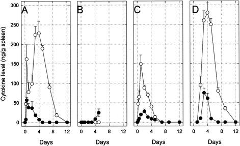
Expression of the proinflammatory cytokines IFN-γ (○) and TNF-α (•) in C57BL/6 mouse spleen following intravenous challenge with 106 pCD− cells of Y. pestis KIM10 (A), 102 pCD+ cells of Y. pestis KIM10 (B), 102 pCD+ cells of Y. pestis KIM10 after the cells were primed with 20 μg of IFN-γ and 20 ng of TNF-α (C), and 102 pCD+ cells of Y. pestis KIM10 by passive immunization on postinfection day 1 with 100 μg of polyclonal rabbit anti-LcrV (D). All untreated mice challenged with pCD+ yersiniae died by postinfection day 6 (B), whereas the remainder survived. This figure is redrawn from the work of Nakajima and Brubaker (67) and Nakajima et al. (68).
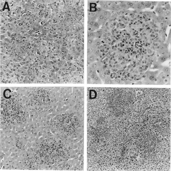
Characteristic histopathological changes in liver caused by Y. pestis KIM10 on postinfection day 3 (hematoxylin and eosin stain). (A) Control mouse infected with pCD+ yersiniae showing multiple necrotic focal lesions without an inflammatory cell response (magnification, ×140); (B) control mouse infected with pCD− yersiniae exhibiting granuloma formation (magnification, ×280); (C) mouse actively immunized with PAV and then infected with pCD+ yersiniae showing protective granulomatous lesions (magnification, ×140); (D) mouse passively immunized with polyclonal rabbit anti-PAV and infected with pCD+ yersiniae showing pregranulomatous lesions prompting infiltration of inflammatory (mononuclear) cells (magnification, ×70). This figure was reprinted from the work of Nakijima et al. (68).
References
-
- Black, D. S., A. Marie-Cardine, B. Schraven, and J. B. Bliska. 2000. The Yersinia tyrosine phosphatase YopH targets a novel adhesion-regulated signalling complex in macrophages. Cell Microbiol. 2:401-414. - PubMed
Publication types
MeSH terms
Substances
LinkOut - more resources
Full Text Sources
Other Literature Sources
