. 2003 Jul;71(7):3979-87.
doi: 10.1128/IAI.71.7.3979-3987.2003.

Borrelia burgdorferi-induced tolerance as a model of persistence via immunosuppression

Affiliations

Borrelia burgdorferi-induced tolerance as a model of persistence via immunosuppression

Isabel Diterich et al. Infect Immun. 2003 Jul.

Abstract

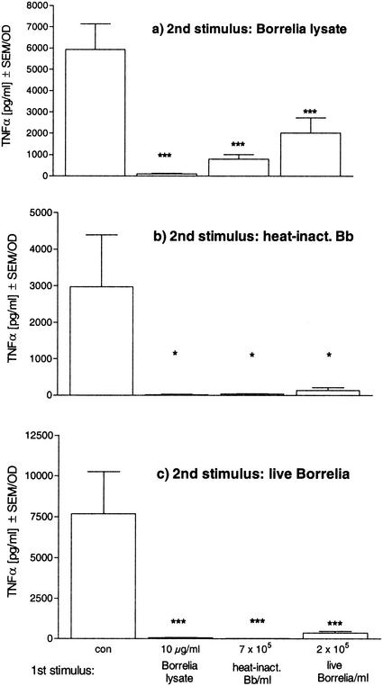
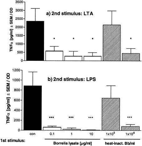
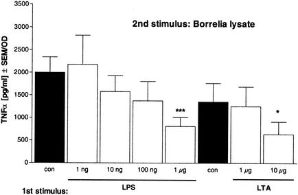
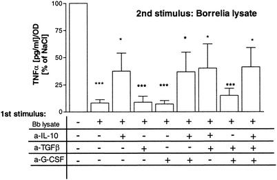
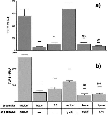
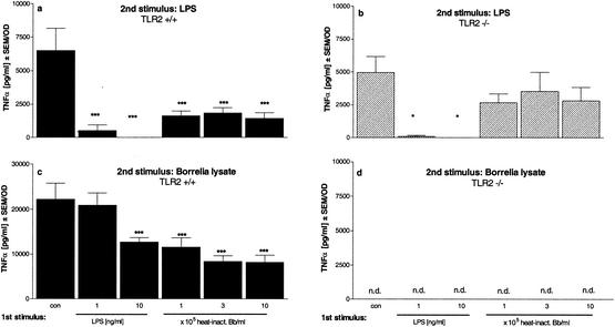
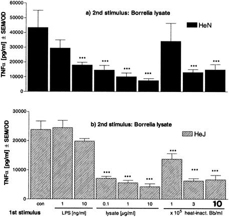
If left untreated, infection with Borrelia burgdorferi sensu lato may lead to chronic Lyme borreliosis. It is still unknown how this pathogen manages to persist in the host in the presence of competent immune cells. It was recently reported that Borrelia suppresses the host's immune response, thus perhaps preventing the elimination of the pathogen (I. Diterich, L. Härter, D. Hassler, A. Wendel, and T. Hartung, Infect. Immun. 69:687-694, 2001). Here, we further characterize Borrelia-induced immunomodulation in order to develop a model of this anergy. We observed that the different Borrelia preparations that we tested, i.e., live, heat-inactivated, and sonicated Borrelia, could desensitize human blood monocytes, as shown by attenuated cytokine release upon restimulation with any of the different preparations. Next, we investigated whether these Borrelia-specific stimuli render monocytes tolerant, i.e. hyporesponsive, towards another Toll-like receptor 2 (TLR2) agonist, such as lipoteichoic acid from gram-positive bacteria, or towards the TLR4 agonist lipopolysaccharide. Cross-tolerance towards all tested stimuli was induced. Furthermore, using primary bone marrow cells from TLR2-deficient mice and from mice with a nonfunctional TLR4 (strain C3H/HeJ), we demonstrated that the TLR2 was required for tolerance induction by Borrelia, and using neutralizing antibodies, we identified interleukin-10 as the key mediator involved. Although peripheral blood mononuclear cells tolerized by Borrelia exhibited reduced TLR2 and TLR4 mRNA levels, the expression of the respective proteins on monocytes was not decreased, ruling out the possibility that tolerance to Borrelia is attributed to a reduced TLR2 expression. In summary, we characterized tolerance induced by B. burgdorferi, describing a model of desensitization which might mirror the immunosuppression recently attributed to the persistence of Borrelia in immunocompetent hosts.


Figures

FIG. 1.
FIG. 1.
Borrelia-induced tolerance in human PBMC. Human PBMC (106) from four healthy donors were incubated with culture medium (con), 10 μg of Borrelia lysate/ml, 7 × 10⁵ heat-inactivated Borrelia cells (heat-inact. Bb), or 2 × 10⁵ live Borrelia cells for 24 h and then washed and incubated for another 24 h in the presence of 10 μg of Borrelia lysate/ml (a), 7 × 10⁵ heat-inactivated Borrelia cells (b), or 2 × 10⁵ live Borrelia cells (c). The concentrations of TNF-α were measured by ELISA. Data are expressed as the means ± SEM divided by the optical density (OD) assessed by the MTT assay. The statistical significance is for comparison with the nontolerized (con) cells. * represents a P value of <0.05 and *** represents a P value of <0.001 based on ANOVA followed by Dunnett's multiple comparison test.
FIG. 2.
FIG. 2.
Induction of Borrelia cross-tolerance to LTA or LPS in human PBMC. Human PBMC (106/well) from four different donors were incubated with culture medium (con, black bars); 0.1, 1, or 10 μg of Borrelia lysate/ml (white bars); or 105 or 106 heat-inactivated Borrelia cells (heat-inact. Bb; striped bars) for 24 h and then washed and incubated for another 24 h in the presence of 10 μg of LTA/ml (a) or 10 ng of LPS/ml (b). The concentrations of TNF-α were measured by ELISA. Data are expressed as the means ± SEM divided by the OD assessed by the MTT test. The statistical significance is for comparison with the nontolerized (con) cells. * represents a P value of <0.05 and *** represents a P value of <0.001 based on ANOVA followed by Dunnett's multiple comparison test.
FIG. 3.
FIG. 3.
Effect of preincubation with LPS or LTA on restimulation with Borrelia lysate. Human PBMC (106/well) were incubated with culture medium (con, black bars); 1, 10, 100, or 1,000 ng of LPS/ml (white bars); or 1 or 10 μg of LTA/ml (white bars) for 24 h and then washed and incubated for another 24 h in the presence of 10 μg of Borrelia lysate/ml. The concentrations of TNF-α were measured by ELISA. Data are expressed as the means ± SEM divided by the OD assessed by the MTT assay. The statistical significance is for comparison with the nontolerized (con) cells. * represents a P value of <0.05 and *** represents a P value of <0.001 based on ANOVA followed by Dunnett's multiple comparison test.
FIG. 4.
FIG. 4.
Neutralization of IL-10, TGF-β, and G-CSF during preincubation prevents establishment of desensitization by Borrelia lysate. PBMC were untreated or were stimulated for 24 h in the presence of Borrelia lysate alone (Bb lysate) or additionally with anti-TGF-β (a-TGFβ; 1 μg/ml) and anti-IL-10 (a-IL-10; 10 μg/ml) monoclonal antibodies, and 1% anti-mu-G-CSF-sheep immunoglobulin G (a-G-CSF) or combinations of the neutralizing antibodies and serum. Controls were cultured without Borrelia lysate. After being washed, cells were restimulated with 10 μg of Borrelia lysate/ml for a further 24 h at 37°C. Cytokine amounts in supernatants were determined by ELISA. Data are expressed as the means ± SEM divided by the OD assessed by the MTT assay. The statistical significance is for comparison with the nontolerized cells. * represents a P value of <0.05 and *** represents a P value of <0.001 based on ANOVA followed by Dunnett's multiple comparison test. +, present; −, absent.
FIG. 5.
FIG. 5.
TLR2 and TLR4 mRNA expression in human PBMC. PBMC (5 × 10⁶) from four different donors were incubated with culture medium, 1 ng of LPS/ml, or 10 μg of Borrelia lysate/ml for 24 h and either lysed or incubated for another 3 h in the presence of medium, 10 μg of Borrelia lysate/ml, or 1 ng of LPS/ml. The cDNA for TLR2 (a), TLR4 (b), and GAPDH (a and b) was amplified, and relative quantification was performed by using a LightCycler. The TLR results were normalized to GAPDH. ** and §§ represent a P value of <0.01 and *** and §§§ represent a P value of <0.001 based on ANOVA followed by Bonferroni's multiple comparison test. *, versus medium and medium; §, versus medium and lysate.
FIG. 6.
FIG. 6.
Induction of tolerance and cross-tolerance in primary bone marrow cells from TLR2+/+ and TLR2−/− mice. Bone marrow cells (5 × 10⁵/well) from TLR2+/+ mice (black bars) and TLR2−/− mice (striped bars) were incubated with culture medium (con), 1 and 10 ng of LPS/ml, or 105, 3 × 10⁵, or 106 heat-inactivated Borrelia cells (heat-inact. Bb) for 24 h and then washed and incubated for another 24 h in the presence of 1 ng of LPS/ml (a and b) or 10 μg of Borrelia lysate/ml (c and d). The concentrations of TNF-α were measured by ELISA. Data are expressed as the means ± SEM divided by the viability assessed by the MTT assay. The statistical significance is for comparison with the nontolerized cells. * represents a P value of <0.05 and *** represents a P value of <0.001 based on ANOVA followed by Dunnett's multiple comparison test. n.d., not detectable.
FIG. 7.
FIG. 7.
Induction of tolerance and cross-tolerance in primary bone marrow cells from C3H/HeN and C3H/HeJ mice. Bone marrow cells (5 × 10⁵/well) from C3H/HeN mice (black bars) and C3H/HeJ mice (striped bars) were incubated with culture medium (con), 1 or 10 ng of LPS/ml, or 105, 3 × 10⁵, or 106 heat-inactivated Borrelia cells (heat-inact. Bb) for 24 h and then washed and incubated for another 24 h in the presence of 10 μg of Borrelia lysate/ml. The concentrations of TNF-α were measured by ELISA. Data are expressed as the means ± SEM divided by the viability assessed by the MTT assay. The statistical significance is for comparison with the nontolerized cells. *** represents a P value of <0.001 based on ANOVA followed by Dunnett's multiple comparison test.

Similar articles

Cited by

References

    1. Aliprantis, A. O., R. B. Yang, M. R. Mark, S. Suggett, B. Devaux, J. D. Radolf, G. R. Klimpel, P. Godowski, and A. Zychlinsky. 1999. Cell activation and apoptosis by bacterial lipoproteins through toll-like receptor-2. Science 285:736-739. - PubMed
    1. Barsig, J., D. S. Bundschuh, T. Hartung, A. Bauhofer, A. Sauer, and A. Wendel. 1996. Control of fecal peritoneal infection in mice by colony-stimulating factors. J. Infect. Dis. 174:790-799. - PubMed
    1. Brightbill, H. D., D. H. Libraty, S. R. Krutzik, R. B. Yang, J. T. Belisle, J. R. Bleharski, M. Maitland, M. V. Norgard, S. E. Plevy, S. T. Smale, P. J. Brennan, B. R. Bloom, P. J. Godowski, and R. L. Modlin. 1999. Host defense mechanisms triggered by microbial lipoproteins through toll-like receptors. Science 285:732-736. - PubMed
    1. Brooke, M. S. 1965. Conversion of immunological paralysis to immunity by endotoxin. Nature 206:635-636. - PubMed
    1. Carroll, J. A., C. F. Garon, and T. G. Schwan. 1999. Effects of environmental pH on membrane proteins in Borrelia burgdorferi. Infect. Immun. 67:3181-3187. - PMC - PubMed

MeSH terms

LinkOut - more resources