. 2003 Jul;71(7):4067-78.
doi: 10.1128/IAI.71.7.4067-4078.2003.

Soluble extracts from Helicobacter pylori induce dome formation in polarized intestinal epithelial monolayers in a laminin-dependent manner

Affiliations

Soluble extracts from Helicobacter pylori induce dome formation in polarized intestinal epithelial monolayers in a laminin-dependent manner

A M Terrés et al. Infect Immun. 2003 Jul.

Abstract

Helicobacter pylori colonizes the stomach at the interface between the mucus layer and the apical pole of gastric epithelial cells. A number of secreted and shed products from the bacteria, such as proteins and lipopolysaccharide, are likely to have a role in the pathogenesis at the epithelial level. To determine the physiological response of transporting polarized epithelia to released soluble factors from the bacterium, we used the T84 cell line. Monolayers of T84 cells were exposed to soluble extracts from H. pylori. The extracts induced rapid "dome" formation as well as an immediate decrease in transepithelial electrical resistance. Domes are fluid-filled blister-like structures unique to polarized epithelia. Their formation has been linked to sodium-transporting events as well as to diminished adherence of the cells to the substrate. H. pylori-induced dome formation in T84 monolayers was exacerbated by amiloride and inhibited by ouabain. Furthermore, it was associated with changes in the expression of the laminin binding alpha 6 beta 4 integrin and the 67-kDa laminin receptor. Domes formed primarily on laminin-coated filters, rather than on fibronectin or collagen matrices, and their formation was inhibited by preincubating the bacterial extract with soluble laminin. This effect was specific to H. pylori and independent of the urease, vacA, cagA, and Lewis phenotype of the strains. These data indicate that released elements from H. pylori can alter the physiological balance and integrity of the epithelium in the absence of an underlying immune response.


Figures
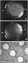
FIG. 1.
FIG. 1.
HPE induce epithelial dome formation. T84 monolayers were grown in semipermeable tissue culture PET inserts and apically stimulated with HPE for 1 h at 37°C. Numerous circular structures appeared in the treated monolayers (A) but not in the controls (B). These structures could be seen with the naked eye. Images shown are from a real-time video recording (A and B) and a phase-contrast image of HPE-treated unfixed cells taken with an inverted microscope (C). Bar = 0.2 mm.
FIG. 2.
FIG. 2.
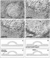
Phase-contrast microscopy of epithelial-matrix, but not cell-cell, detachment in HPE-treated monolayers. (A) Magnified dome as seen with an inverted phase-contrast microscope. Domes correspond to areas in which the monolayer is detached from the substrate. (B to D) Sequence of images of the monolayer taken as it is focused from the bottom to the top of the dome. The diagrams represent a transverse section of panels A to D. Arrows in the diagrams correspond to the asterisks in the photographs. Sequential focusing shows the porous support (B), the attached monolayer above it (C), and then the detached cells at the top of the dome (D). Bar = 300 μm.
FIG. 3.
FIG. 3.
Dome formation is accompanied by a decrease in TER and is inhibited by soluble laminin. Monolayers were grown in PET semipermeable inserts and stimulated with HPE for 1 h. (A and C) TER was measured with an Endhom electrode and is represented as the percentage of control values at the beginning of the experiment. (B to D) Epithelial detachment was quantified by the number of domes and is represented as a percentage of the positive control (monolayer apically treated with HPE for 1 h) of every experiment. HPE-treated monolayers had a reduced TER (A), independent of the nature of the substrate on which they were grown (PET or polyester plastic, laminin, fibronectin, or collagen) (C). However, they primarily developed domes when grown on PET or laminin (D) and only when they were apically, but not basally, stimulated with HPE (B). (E) The effect of soluble laminin in HPE-induced dome formation was subsequently assessed. HPE was preincubated for 10 min with three concentrations of soluble laminin as indicated and then used to apically stimulate the cells for comparison with untreated HPE. (F) Alternatively, monolayers were treated with 10 μg of soluble laminin (L) diluted in tissue culture medium for 30 min, then washed and incubated with HPE, and compared to monolayers only treated with HPE and without laminin preincubation (HPE).
FIG. 4.
FIG. 4.
Dome formation is reversible and only occurs in transporting epithelia. Cells were grown and analyzed as indicated in the legend for Fig. 3. (A and B) The effect of monolayer age on HPE-induced TER decrease and dome formation was assessed. (C) HPE-induced dome formation was also assessed in different cell lines, as indicated. (D) Reversibility of dome formation and TER decrease was assessed by replacing HPE with normal tissue culture medium (arrow) and culturing the cells overnight.
FIG. 5.
FIG. 5.
HPE specifically induces dome formation in a concentration-dependent manner and is independent of the urease, VacA, CagA, and Lewis x and y phenotypes of the strains. (A) The capacity to induce domes was tested with extracts from H. pylori strains of defined phenotype (26). NUD, nonulcer dyspepsia; DU, duodenal ulcer; Rex, reflux esophagitis; Rs, reference strain; UTI, urinary tract infection; Le x, Lewis x; Le y, Lewis y. (B) Dome formation was assessed with extracts from H. pylori reference strains NCTC 11637 and 11638, as well as clinical isolates MI 215, 229, 300, and 270. Extracts from E. coli (wt = wild type clinical isolate; DH5α = cloning vector) and S. marcescens were used for comparative purposes. (C) Monolayer stimulation with serial dilutions of HPE from NCTC 11638 showed that dome formation was concentration dependent. The number of domes (detachment) is expressed as a percentage of the number of domes induced by undiluted HPE from strain NCTC 11638. (D) The specific involvement of urease, VacA, and CagA in dome formation was assessed by stimulating T84 monolayers with HPE from parental wild-type strains (84183, 60190, and N6) and the corresponding isogenic mutants for vacA, cagA, ureB, and ureG under kanamycin (km) selection. Normalization between strains (B and D) was achieved by adjusting the bacterial solution to an optical density at 600 nm of 2.9 before removing cells by centrifugation.
FIG. 6.
FIG. 6.
Ultrastructural analysis of HPE-treated and control monolayers by transmission electron microscopy. Ultrastructural analysis by transmission electron microscopy was performed in control (A, C, and E) and treated (B, D, and F) monolayers. A full-width monolayer (A and B), apical domain (C and D), and basolateral domain (E and F) are represented. Bars: 5 μm (A and B), 0.5 μm (C and D), and 1 μm (E and F). n, nuclei; bl sp, basolateral space; tj, tight junctions.
FIG. 7.
FIG. 7.
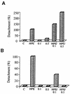
Dome formation is inhibited by ouabain but exacerbated by amiloride. The effect of amiloride (A) and ouabain (B) on HPE-induced dome formation was assessed. Monolayers were pretreated with 0.1 and 0.5 mM amiloride (apical) or ouabain (basal) for 30 min prior to incubation with HPE for a further hour (HPE/0.1 and HPE/0.5) and compared to untreated controls (C), HPE-treated cells (HPE), and cells treated with the same concentrations of amiloride or ouabain alone (0.1 and 0.5).
FIG. 8.
FIG. 8.
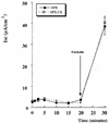
Dome formation is not associated with chloride secretion. T84 intestinal epithelial monolayers were examined for induction of electrogenic Cl secretion by HPE. Shown here are time course curves of Cl secretion (measured as Isc) for HPE and control. To demonstrate that cells were capable of Isc, the cAMP-mediated agonist forskolin (3 μM) was added at 20 min. TER and Isc were measured as indicated in Materials and Methods. Data were pooled from six to nine individual monolayers for each condition, and results are expressed as the mean peak Isc ± the standard error of the mean.
FIG. 9.
FIG. 9.
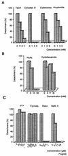
Dome formation is sensitive to cytoskeletal disruption and inhibitors of phosphorylation-mediated signaling pathways. Inhibitors and disrupters were added to the cells for 30 min prior to incubation with HPE for a further hour; detachment was quantified by counting the number of domes and is represented as a percentage of the positive control treated with HPE alone within each experiment. Experiments were performed in individual inserts and repeated three separate times. The graph represents the results from a typical experiment. Cytochal. D, cytochalasin D; NaAz, sodium azide; PT, pertussis toxin; cyclosp., cyclosporine; Staur., staurosporine; Herb. A, herbimycin A.
FIG. 10.
FIG. 10.
Modulation of basolateral epithelial integrin distribution upon exposure to HPE. Immunofluorescence staining of control (A, C, E, and G) and HPE-treated (B, D, F, and H) monolayers for β1 integrin (A and B), α6 integrin (C and D), β4 integrin (E and F), and the 67LR (G and H). T84 polarized monolayers grown in 1-cm2 PET tissue culture inserts were apically stimulated for 1 h with HPE. Control monolayers were maintained in medium alone. The monolayers were subsequently fixed and stained by indirect immunofluorescence with anti-β1, anti-β4, GoH3, and MluC5 antibodies and analyzed under an inverted fluorescence microscope. Bar = 30 μm.

Similar articles

Cited by

References

    1. Ardini, E., E. Tagliabue, A. Magnifico, S. Buto, V. Castronovo, M. I. Colhaghi, and S. Menard. 1997. Co-regulation and physical association of the 67-kDa monomeric laminin receptor and the α6β4 integrin. J. Biol. Chem. 272:2342-2345. - PubMed
    1. Ashorn, M., F. Cantet, K. Mayo, and F. Megraud. 2000. Cytoskeletal rearrangements induced by Helicobacter pylori strains in epithelial cell culture: possible role of the cytotoxin. Dig. Dis. Sci. 45:1774-1780. - PubMed
    1. Awayda, M. 2000. Specific and nonspecific effects of protein kinase C on the epithelial Na+ channel. J. Gen. Physiol. 115:559-570. - PMC - PubMed
    1. Barrett, K. 2000. New insights into the pathogenesis of intestinal dysfunction: secretory diarrhea and cystic fibrosis. World J. Gastroenterol. 6:470-474. - PMC - PubMed
    1. Bradford, M. M. 1976. A rapid and sensitive method for the quantitation of microgram quantities of protein utilizing the principle of protein-dye binding. Anal. Biochem. 72:248-254. - PubMed

Publication types

MeSH terms

LinkOut - more resources