. 2003 Jun;13(6B):1466-77.
doi: 10.1101/gr.1087603.

G protein-coupled receptor genes in the FANTOM2 database

Affiliations

G protein-coupled receptor genes in the FANTOM2 database

Yuka Kawasawa et al. Genome Res. 2003 Jun.

Abstract

G protein-coupled receptors (GPCRs) comprise the largest family of receptor proteins in mammals and play important roles in many physiological and pathological processes. Gene expression of GPCRs is temporally and spatially regulated, and many splicing variants are also described. In many instances, different expression profiles of GPCR gene are accountable for the changes of its biological function. Therefore, it is intriguing to assess the complexity of the transcriptome of GPCRs in various mammalian organs. In this study, we took advantage of the FANTOM2 (Functional Annotation Meeting of Mouse cDNA 2) project, which aimed to collect full-length cDNAs inclusively from mouse tissues, and found 410 candidate GPCR cDNAs. Clustering of these clones into transcriptional units (TUs) reduced this number to 213. Out of these, 165 genes were represented within the known 308 GPCRs in the Mouse Genome Informatics (MGI) resource. The remaining 48 genes were new to mouse, and 14 of them had no clear mammalian ortholog. To dissect the detailed characteristics of each transcript, tissue distribution pattern and alternative splicing were also ascertained. We found many splicing variants of GPCRs that may have a relevance to disease occurrence. In addition, the difficulty in cloning tissue-specific and infrequently transcribed GPCRs is discussed further.


Figures
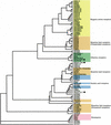
Figure 1
Figure 1
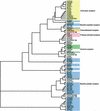
Classification tree (Family A—small molecule). A rooted tree was constructed for 83 GPCRs. GPCRs that have cognate ligands are distinguished in colored subgroups. Orphan GPCRs are shown in uncolored branches, and novel genes are indicated with black circles. Abbreviations are shown in Supplementary Information 1 (available online at www.genome.org).
Figure 2
Figure 2
Classification tree (Family A—peptide). A rooted tree was constructed for 82 GPCRs. GPCRs that have cognate ligands are distinguished in colored subgroups. Orphan GPCRs are shown in uncolored branches, and novel genes are indicated with black circles. Abbreviations are shown in Supplementary Information 2.
Figure 4
Figure 4
Classification tree (Family C). An unrooted tree was constructed for 11 GPCRs. The scale bar indicates a maximum likelihood branch length of 0.1 inferred substitutions per site. GPCRs that have cognate ligands are distinguished in colored subgroups. Orphan GPCRs are shown in uncolored branches, and novel genes are indicated with black circles. Abbreviations are shown in Supplementary Information 4.
Figure 3
Figure 3
Classification tree (Family B). An unrooted tree was constructed for 24 GPCRs. The scale bar indicates a maximum likelihood branch length of 0.1 inferred substitutions per site. GPCRs that have cognate ligands are distinguished in colored subgroups. Orphan GPCRs are shown in uncolored branches. Abbreviations are shown in Supplementary Information 3.
Figure 5
Figure 5
Predicted splicing variants of Gpr83. Schematic representation of the mouse GPR83 polypeptide and splicing alternatives generating the different variants. Chromosomal localization was obtained by genome mapping. NM_010287 is the Gpr83 gene registered in the public database, and blue bars represent each exon of Gpr83. Four RIKEN clones (9530022I23, C030041M14, A630019F13, and 5330401A04) were mapped against the Gpr83 gene; red bars represent the predicted coding region of each RIKEN clone. Edited mRNA and predicted ORF sequences are also illustrated. The seven transmembrane (7TM) region is shown in black bar. Green bars show the predicted coding regions that do not match data in the public database. Variants RP23 and RP39 have been described previously (Harrigan et al. 1991).
Figure 6
Figure 6
Predicted variants of Gpr37. Schematic representation of the mouse GPR37 polypeptide and splicing alternatives that generate the different variants. Chromosomal localization was obtained by genome mapping. NM_010338 is the Gpr37 gene registered in the public database; blue bars represent each exon of Gpr37. Three RIKEN clones (6430580C01, E130007J18, and A930017K23) were mapped against the Gpr37 gene; red bars represent the predicted coding region of each RIKEN clone. Edited mRNA and predicted ORF sequences are also illustrated. The seven transmembrane (7TM) region is shown in black bar. Green bars show the predicted coding regions that do not match data in the public database. Clone E130007J18 is predicted to lack the precedent region of exon 1, and A930017K23 is predicted to have a shorter exon 2.

References

    1. Akhundova, A., Getmanova, E., Gorbulev, V., Carnazzi, E., Eggena, P., and Fahrenholz, F. 1996. Cloning and functional characterization of the amphibian mesotocin receptor, a member of the oxytocin/vasopressin receptor superfamily. Eur. J. Biochem. 237: 759-767. - PubMed
    1. Alcedo, J., Ayzenzon, M., Von Ohlen, T., Noll, M., and Hooper, J.E. 1996. The Drosophila smoothened gene encodes a seven-pass membrane protein, a putative receptor for the hedgehog signal. Cell 86: 221-232. - PubMed
    1. Ashburner, M., Ball, C.A., Blake, J.A., Botstein, D., Butler, H., Cherry, J.M., Davis, A.P., Dolinski, K., Dwight, S.S., Eppig, J.T., et al. 2000. Gene ontology: Tool for the unification of biology. The Gene Ontology Consortium. Nat. Genet. 25: 25-29. - PMC - PubMed
    1. Baldarelli, R.M., Hill, D.P., Blake, J.A., Adachi, J., Furuno, M., Bradt, D., Corbani, L.E., Cousins, S., Frazer, K.S., Qi, D., et al. 2003. Connecting sequence and biology in the laboratory mouse. Genome Res. (this issue). - PMC - PubMed
    1. Bartfai, T., Hokfelt, T., and Langel, U. 1993. Galanin—A neuroendocrine peptide. Crit. Rev. Neurobiol. 7: 229-274. - PubMed

WEB SITE REFERENCES

    1. ftp://ftp.ncbi.nih.gov/genbank/genomes/M_musculus/CHR_Y/; mouse Y-chromosome.
    1. ftp://wolfram.wi.mit.edu/pub/mouse_contigs/MGSC_V3; the MGSCv3 assembly.
    1. http://genome.cse.ucsc.edu/goldenPath/22Dec2001; human Y-chromosome sequences, GoldenPath.
    1. http://genomes.rockefeller.edu/MouSDB; M. Zavolan comprehensive database of probable splice variants.
    1. http://www.informatics.jax.org; MGI: Mouse Genome Informatics resource.

Publication types

MeSH terms

Substances