. 2003 Spring;8(1):37-52.
doi: 10.1379/1466-1268(2003)8<37:ivrfva>2.0.co;2.

Integrin alphavbeta3, requirement for VEGFR2-mediated activation of SAPK2/p38 and for Hsp90-dependent phosphorylation of focal adhesion kinase in endothelial cells activated by VEGF

Affiliations

Integrin alphavbeta3, requirement for VEGFR2-mediated activation of SAPK2/p38 and for Hsp90-dependent phosphorylation of focal adhesion kinase in endothelial cells activated by VEGF

Bénédicte Masson-Gadais et al. Cell Stress Chaperones. 2003 Spring.

Abstract

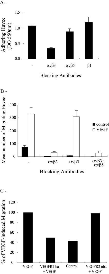
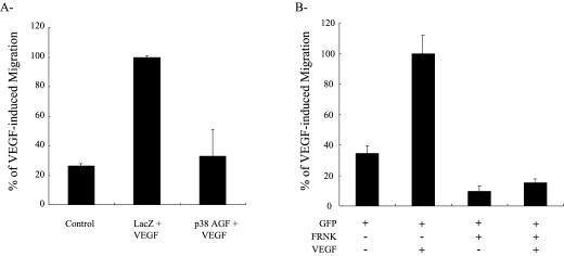
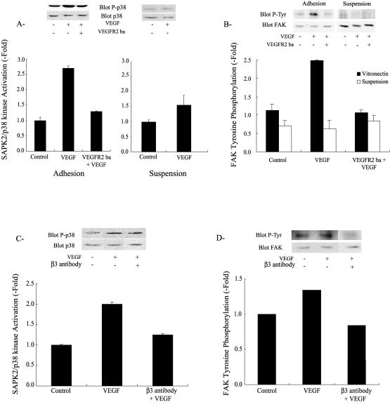
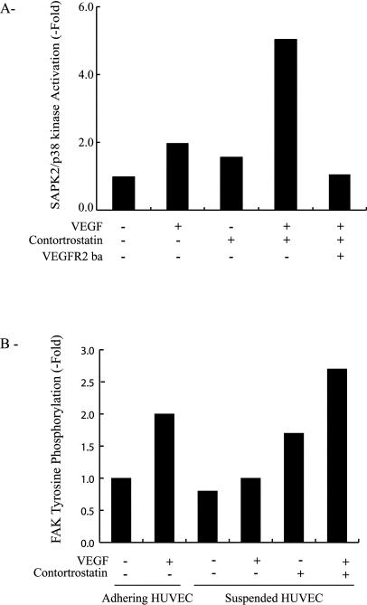
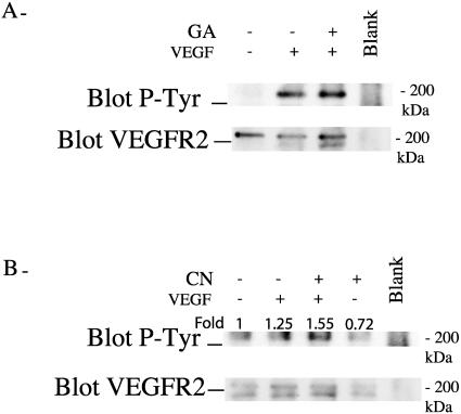
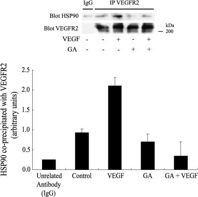
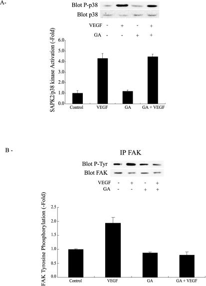
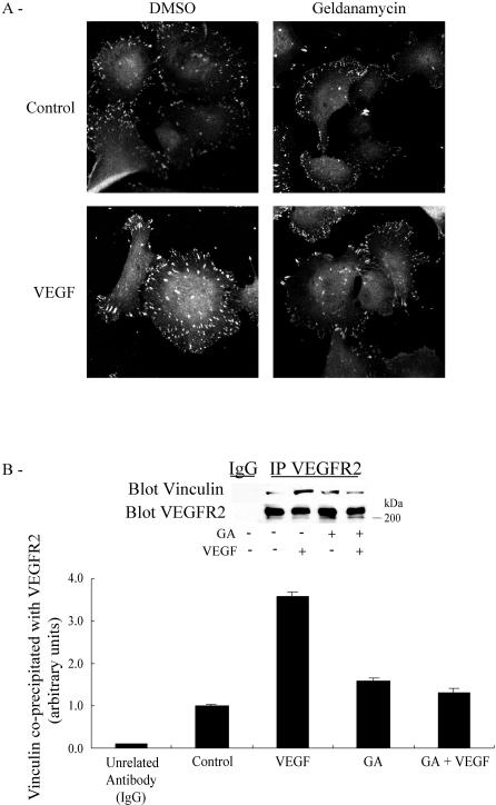
Endothelial cell migration, a key process in angiogenesis, requires the coordinated integration of motogenic signals elicited by the adhesion of endothelial cells to extracellular matrices and by angiogenic cytokines such as the vascular endothelial growth factor (VEGF). In this study, we found that addition of VEGF to human umbilical vein endothelial cells cultivated on vitronectin triggers a synergistic interaction between the VEGF receptor VEGFR2 and the clustered integrin receptor alphavbeta3. The interaction between VEGFR2 and alphavbeta3 is required for full phosphorylation of VEGFR2 and to drive the activation of motogenic pathways involving focal adhesion kinase (FAK) and stress-activated protein kinase-2/p38 (SAPK2/p38). The signal emanating from the VEGFR2 and alphavbeta3 interaction and leading to SAPK2/p38 activation proceeds directly from VEGFR2. The chaperone Hsp90 is found in a complex that coprecipitates with inactivated VEGFR2, and the association is increased by VEGF and decreased by geldanamycin, a specific inhibitor of Hsp90-mediated events. Geldanamycin also impairs the phosphorylation of FAK that results from the interaction between VEGFR2 and alphavbeta3, and this is accompanied by an inhibition of the recruitment of vinculin to VEGFR2. We conclude that a necessary cross talk should occur between VEGFR2 and the integrin alphavbeta3, to transduce the VEGF signals to SAPK2/p38 and FAK and that Hsp90 is instrumental in the building up of focal adhesions by allowing the phosphorylation of FAK and the recruitment of vinculin to VEGFR2.


Figures

Fig 1.
Fig 1.
VEGF-induced migration of HUVEC on vitronectin is mediated by VEGFR2 and integrin αvβ3. (A) HUVEC in suspension were either not incubated or incubated for 20 minutes with optimal concentrations or dilutions of anti-αvβ3 (1 μg/mL), anti-αvβ5 (1 μg/mL), or anti-β1 (1:1000) integrin antibodies. Cells were then plated in 96-well culture dishes precoated with 3 μg/mL vitronectin. After 60 minutes at 37°C, adhering cells were stained with crystal violet and quantified by optical density (550 nm). Data points represent means ± SD of triplicate samples from 3 different experiments. (B) HUVEC in suspension were either not preincubated or preincubated for 20 minutes with anti-αvβ3 (1 μg/mL), anti-αvβ5 (1 μg/mL), or anti-β1 (1:1000) integrin antibodies. (C) HUVEC in suspension were preincubated in the presence or absence of a VEGFR2 blocking (VEGFR2-ba 10 μg/mL, 20 minutes) or nonblocking antibody (VEGFR2-nba 10 μg/mL, 20 minutes). After treatments, in both B and C, cells were seeded on the upper part of a vitronectin-coated membrane in a modified Boyden chamber either containing or not containing VEGF (5 ng/mL) in the lower part. After 4 hours of migration, cells on the upper part of the membrane were scraped, and the cells on the lower part were stained with Mayer's hematoxylin. The cells of each well were counted in 5 fields at 100× magnification. Data points represent means from triplicate samples taken from at least 3 different experiments. VEGF, vascular endothelial growth factor; HUVEC, human umbilical vein endothelial cells; VEGFR2, VEGF receptor 2
Fig 2.
Fig 2.
VEGF-induced migration of HUVEC on vitronectin is mediated by activation of SAPK2/p38 and FAK. (A) HUVEC were infected with adenoviral vectors expressing Lacz or p38 AGF, a dominant negative form of p38α. (B) HUVEC were electroporated at 25 μF and 300 V with a total of 30 μg of a GFP construct with or without a FRNK construct. In both A and B, infected and transfected cells were seeded on the upper part of a vitronectin-coated membrane in a modified Boyden chamber either containing or not containing VEGF (5 ng/mL) in the lower part. After 4 hours of migration, cells on the upper part of the membrane were scraped. In A, the cells on the lower part were stained with Mayer's hematoxylin and counted in 5 fields at 100× magnification. In B, fluorescent cells, on the lower part, expressing FRNK or GFP (or both) were counted in 7 fields using an inverted fluorescence microscope at 200×. Data points represent means of triplicate samples. VEGF, vascular endothelial growth factor; HUVEC, human umbilical vein endothelial cells; SAPK2/p38, stress-activated protein kinase-2/p38; FAK, focal adhesion kinase; GFP, green fluorescent protein; FRNK, FAK-related nonkinase protein
Fig 3.
Fig 3.
VEGF-induced SAPK2/p38 activation and FAK tyrosine phosphorylation require both VEGFR2 and integrin αvβ3. (A) Quiescent HUVEC on vitronectin or in suspension were either not pretreated or pretreated with a VEGFR2 blocking antibody (VEGFR2-ba 10 μg/mL, 20 minutes) and then were either treated or not treated with VEGF (5 ng/mL, 5 minutes). Cells were then extracted and subjected to SAPK2/p38 assay. Extracts were separated by SDS-PAGE and transferred to a nitrocellulose membrane. The membrane was processed by Western blot for phospho p38 detection. The membrane was stripped and reprobed for total p38 to ensure equal protein loading. Data points represent means ± SD of duplicate samples from 2 separate experiments. Representative blots are shown. (B) Quiescent HUVEC were maintained on vitronectin or put in suspension for 20 minutes. Adhering cells were either not pretreated or pretreated with a VEGFR2 blocking antibody (VEGFR2-ba 10 μg/mL, 20 minutes) and then were treated with VEGF (5 ng/mL, 5 minutes). Cells in suspension were either not pretreated or pretreated with the VEGFR2 blocking antibody (VEGFR2-ba 10 μg/mL, 20 minutes), and VEGF (5 ng/mL) was added for the last 5 minutes of suspension. Cells were then extracted and subjected to FAK immunoprecipitation, separated on SDS-PAGE, and transferred to a nitrocellulose membrane. The membrane was processed for phosphotyrosine detection. The membrane was stripped and reprobed for total FAK to ensure equal protein loading. Data points represent means ± SD of duplicate samples from 2 separate experiments. Representative blots are shown. (C, D) Cells in suspension were either not pretreated or pretreated with anti-integrin αvβ3 antibody (1 μg/mL, 20 minutes) and then were plated on vitronectin for 60 minutes. After 55 minutes, cells were either not treated or treated with VEGF (5 ng/mL, 5 minutes). In C, cells were extracted and subjected to p38 assay as in A. In D, cells were extracted and subjected to FAK assay as in B. Data points represent means of triplicate samples. Representative blots are shown. VEGF, vascular endothelial growth factor; SAPK2/p38, stress-activated protein kinase-2/p38; FAK, focal adhesion kinase; HUVEC, human umbilical vein endothelial cells; VEGFR2, VEGF receptor 2; SDS-PAGE, sodium dodecyl sulfate–polyacrylamide gel electrophoresis
Fig 4.
Fig 4.
VEGF-induced SAPK2/p38 activation and FAK tyrosine phosphorylation require clustering of integrin αvβ3. (A) Quiescent cells were put in suspension and were either not pretreated or pretreated for the last 10 minutes of suspension with 10 nM contortrostatin. Then, they were either not treated or treated with VEGF (5 ng/mL, last 5 minutes of suspension) in the presence or absence of the VEGFR2 blocking antibody (VEGFR2-ba 10 μg/mL). Cells were then extracted and processed for SAPK2/p38 assay as in Figure 3A. Data points represent means of triplicate samples for each condition. Results are representative of 2 different experiments. (B) Quiescent HUVEC maintained on vitronectin were left to adhere or were put in suspension for 20 minutes. Adhering cells were either not treated or treated with VEGF (5 ng/mL, 5 minutes). Cells in suspension were either not pretreated or pretreated with 10 nM contortrostatin for the last 10 minutes of suspension and were either not treated or treated with VEGF (5 ng/mL, last 5 minutes of suspension). Cells were then extracted and subjected to FAK phosphorylation assay as in Figure 3B. Data points represent means of duplicate samples. Results are representative of 2 different experiments. VEGF, vascular endothelial growth factor; SAPK2/p38, stress-activated protein kinase-2/p38; FAK, focal adhesion kinase; VEGFR2, VEGF receptor 2; HUVEC, human umbilical vein endothelial cells
Fig 5.
Fig 5.
Integrin αvβ3 mediates tyrosine phosphorylation of VEGFR2 in response to VEGF. (A, B) Quiescent HUVEC were maintained on vitronectin or put in suspension for 20 minutes. Adhering cells (A) were either not pretreated or pretreated with geldanamycin (GA, 1 μg/mL, 60 minutes) and then were either not treated or treated with VEGF (5 ng/mL, 5 minutes). For cells in suspension (B), contortrostatin (CN, 10 nM) was either not added or added for the last 10 minutes of suspension, and VEGF (5 ng/mL) was either not added or added for the last 5 minutes. Cells were then extracted and subjected to VEGFR2 immunoprecipitation. Thereafter, they were separated on sodium dodecyl sulfate–polyacrylamide gel electrophoresis and transferred to a nitrocellulose membrane to detect phosphotyrosinated VEGFR2. The membrane was stripped and reprobed for total VEGFR2 to ensure equal protein loading. In the blank track (A, B), no cellular extract was added. Results are representative of 2 different experiments. VEGF, vascular endothelial growth factor; VEGFR2, VEGF receptor 2; HUVEC, human umbilical vein endothelial cells
Fig 6.
Fig 6.
Hsp90 coprecipitates with VEGFR2. Quiescent HUVEC maintained on vitronectin were either not pretreated or pretreated for 60 minutes with geldanamycin (1 μg/mL) and then they were either not treated or treated with VEGF (5 ng/mL, 5 minutes). Cells were then extracted and subjected to VEGFR2 immunoprecipitation and were processed for immunodetection of Hsp90, as described in Materials and Methods. Data points are means ± SD of duplicate samples from 2 different experiments. Representative blots are shown. VEGF, vascular endothelial growth factor; VEGFR2, VEGF receptor 2; HUVEC, human umbilical vein endothelial cells
Fig 7.
Fig 7.
Geldanamycin does not inhibit SAPK2/p38 activation induced by VEGF, but it does block tyrosine phosphorylation of FAK. (A, B) Quiescent HUVEC on vitronectin were either not pretreated or pretreated for 60 minutes with geldanamycin (1 μg/mL) and then they were subsequently either not treated or treated with VEGF (5 ng/mL, 5 minutes). Cells were then extracted, separated on sodium dodecyl sulfate–polyacrylamide gel electrophoresis, and SAPK2/p38 (A) and tyrosine phosphorylation of FAK (B) were assessed as described in Figure 3. Data points represent means ± SD of triplicate samples from 2 different experiments. Representative blots are shown. SAPK2/p38, stress-activated protein kinase-2/p38; VEGF, vascular endothelial growth factor; FAK, focal adhesion kinase; HUVEC, human umbilical vein endothelial cells
Fig 8.
Fig 8.
VEGF-mediated recruitment of vinculin to VEGFR2 and focal adhesion assembly are inhibited by geldanamycin. (A) Quiescent HUVEC plated on vitronectin-coated labtek chamber were either not pretreated or pretreated with geldanamycin (1 μg/mL, 60 minutes) and then were either not exposed or exposed for 15 minutes to VEGF (5 ng/mL). Cells were fixed, and vinculin was detected with vinculin antibody and then revealed with anti-mouse IgG coupled to Alexa 568. Cells were examined by fluorescence microscopy. Representative fields are shown. (B) Quiescent HUVEC on vitronectin were either not pretreated or pretreated for 60 minutes with geldanamycin (1 μg/mL), and they were subsequently either treated or treated with VEGF (5 ng/mL, 5 minutes). Cells were then extracted and subjected to VEGFR2 immunoprecipitation, separated on sodium dodecyl sulfate–polyacrylamide gel electrophoresis, and transferred to a nitrocellulose membrane. The membrane was then processed by Western blot for immunodetection of vinculin. Data points represent means ± SD of triplicate samples from 3 different experiments. Representative blots are shown. VEGF, vascular endothelial growth factor; VEGFR2, VEGF receptor 2; HUVEC, human umbilical vein endothelial cells

Similar articles

Cited by

References

    1. Aplin AE, Howe A, Alahari SK, Juliano RL. Signal transduction and signal modulation by cell adhesion receptors: the role of integrins, cadherins, immunoglobulin-cell adhesion molecules, and selectins. Pharmacol Rev. 1998;50:197–263. - PubMed
    1. Borges E, Jan Y, Ruoslahti E. PDGF-receptor-(beta) and EGF-receptor-2 bind to the (beta)3 integrin through its extracellular domain. J Biol Chem. 2000;275:39867–39873. - PubMed
    1. Byzova TV, Goldman CK, Pampori N, Thomas KA, Bett A, Shattil SJ, Plow EF. A mechanism for modulation of cellular responses to VEGF: activation of the integrins. Mol Cell. 2000;6:851–860. - PubMed
    1. Chouinard N, Valerie K, Rouabhia M, Huot J. UVB-mediated activation of p38 mitogen-activated protein kinase enhances resistance of normal human keratinocytes to apoptosis by stabilizing p53. Biochem J. 2002;365:133–145. - PMC - PubMed
    1. Dedhar S, Williams B, Hannigan G. Integrin-linked kinase (ILK): a regulator of integrin and growth-factor signalling. Trends Cell Biol. 1999;9:319–323. - PubMed

Publication types

MeSH terms