. 2003 Jun 23;161(6):1151-61.
doi: 10.1083/jcb.200212158.

Critical role for scaffolding adapter Gab2 in Fc gamma R-mediated phagocytosis

Affiliations

Critical role for scaffolding adapter Gab2 in Fc gamma R-mediated phagocytosis

Haihua Gu et al. J Cell Biol. .

Abstract

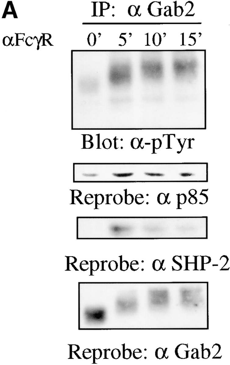
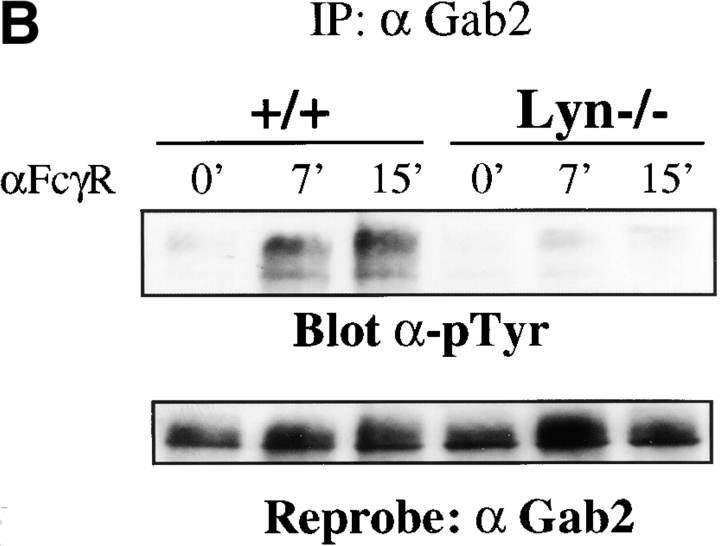
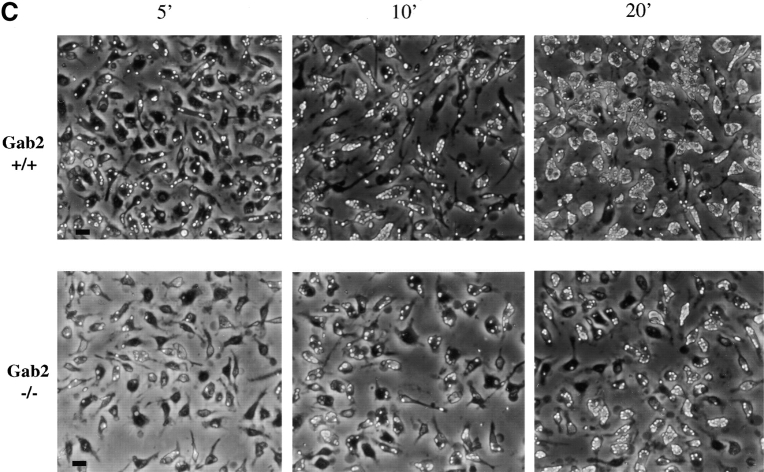
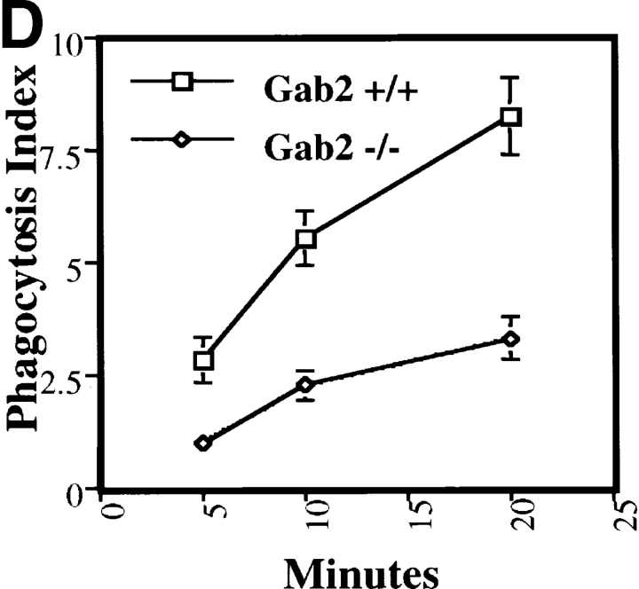
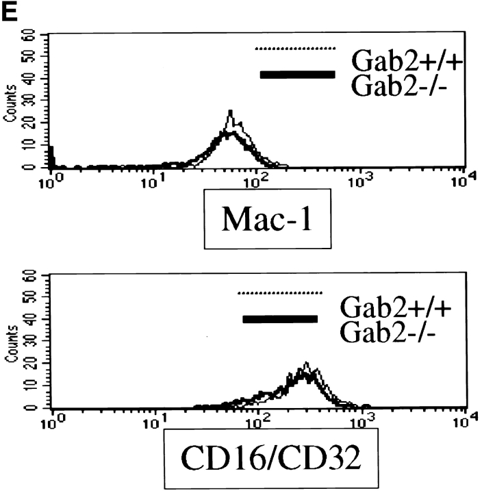
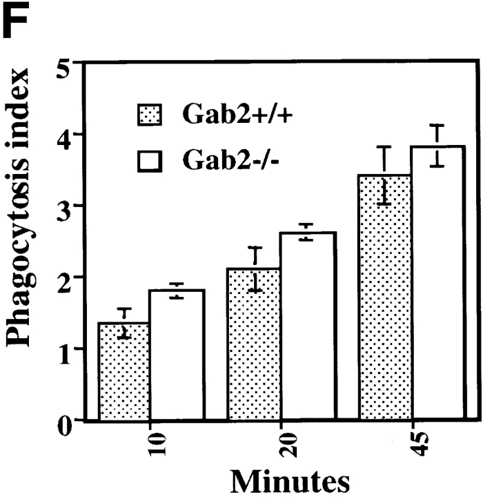
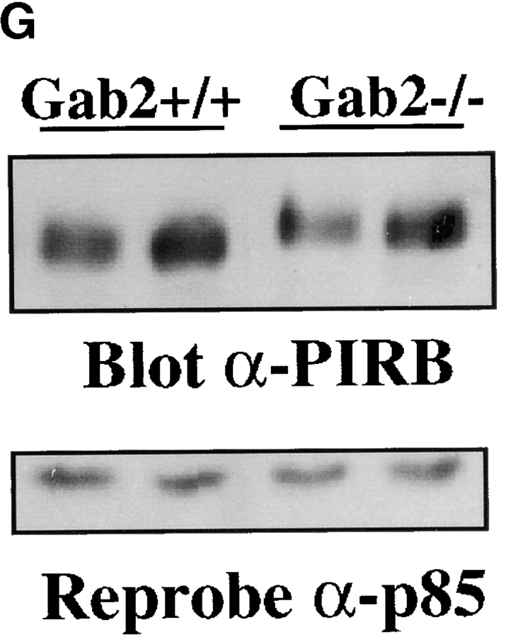
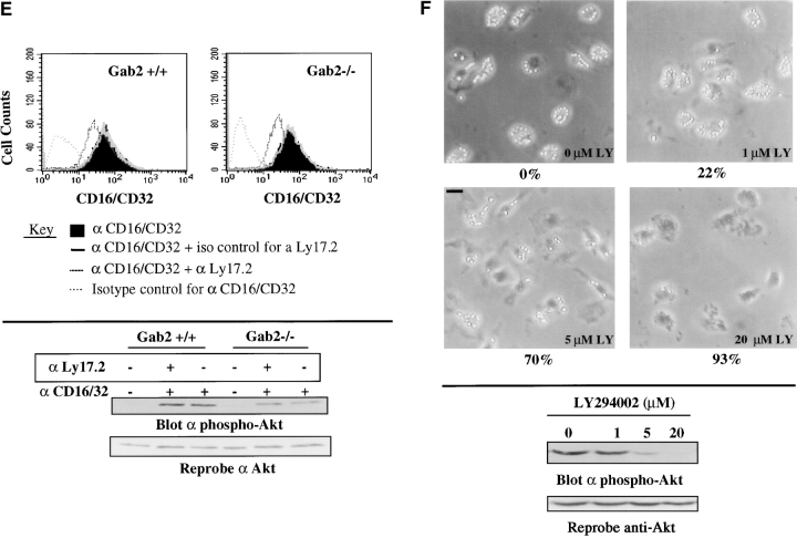
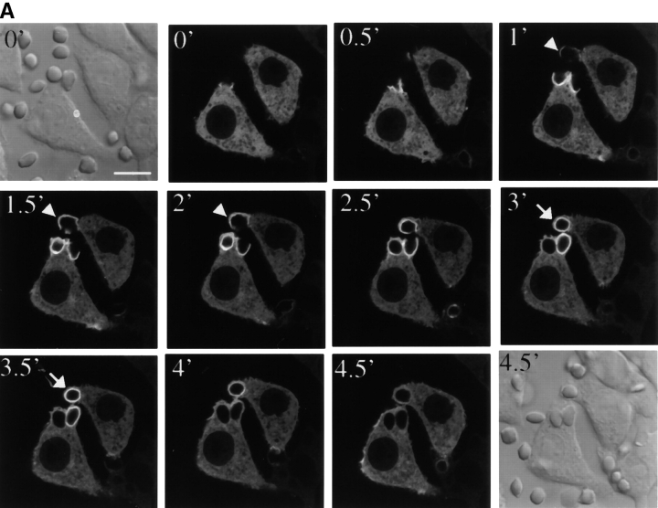
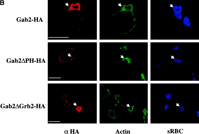
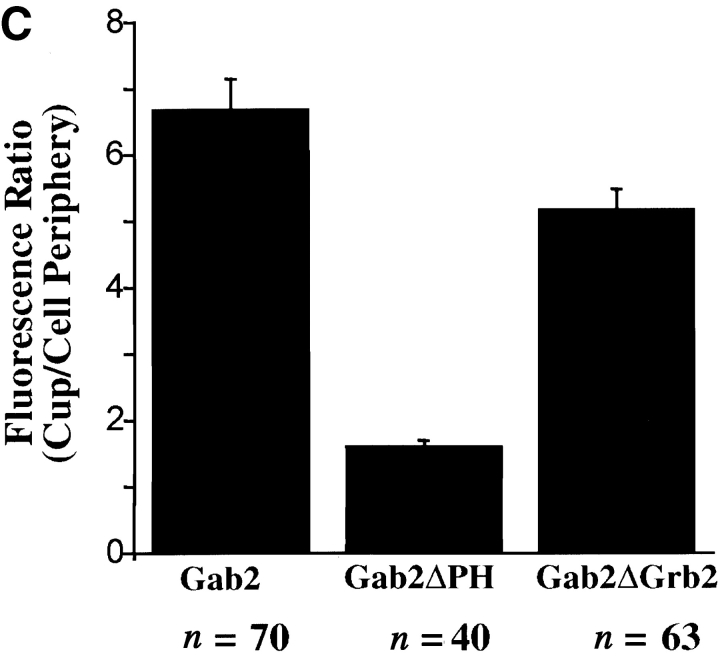
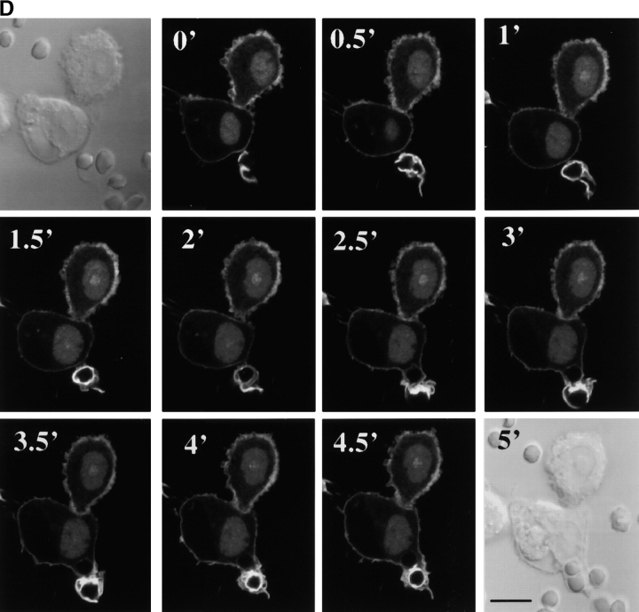
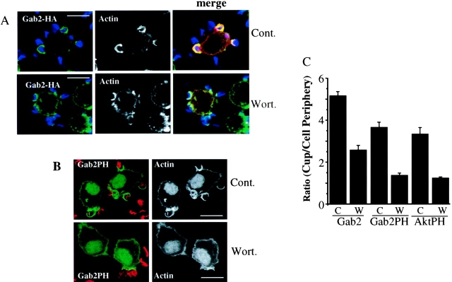
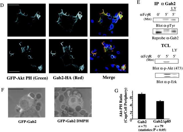
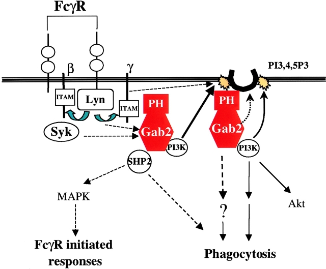
Grb2-associated binder 2 (Gab2), a member of the Dos/Gab subfamily scaffolding molecules, plays important roles in regulating the growth, differentiation, and function of many hematopoietic cell types. In this paper, we reveal a novel function of Gab2 in Fcgamma receptor (FcgammaR)-initiated phagocytosis in macrophages. Upon FcgammaR activation, Gab2 becomes tyrosyl phosphorylated and associated with p85, the regulatory subunit of phosphoinositide 3-kinase (PI3K), and the protein-tyrosine phosphatidylinositol Shp-2. FcgammaR-mediated phagocytosis is severely impaired in bone marrow-derived macrophages from Gab2-/- mice. The defect in phagocytosis correlates with decreased FcgammaR-evoked activation of Akt, a downstream target of PI3K. Using confocal fluorescence microscopy, we find that Gab2 is recruited to the nascent phagosome, where de novo PI3K lipid production occurs. Gab2 recruitment requires the pleckstrin homology domain of Gab2 and is sensitive to treatment with the PI3K inhibitor wortmannin. The Grb2 binding site on Gab2 also plays an auxiliary role in recruitment to the phagosome. Because PI3K activity is required for FcgammaR-mediated phagocytosis, our results indicate that Gab2 acts as a key component of FcgammaR-mediated phagocytosis, most likely by amplifying PI3K signaling in the nascent phagosome.


Figures

Figure 1.
Figure 1.
Gab2 is critical for FcγR-mediated phagocytosis in BMM. (A) FcγR cross-linking evokes Gab2 tyrosyl phosphorylation and complex formation. BMM from a wild-type (+/+) mouse were starved and stimulated by FcγR cross-linking for the indicated times. Lysates were immunoprecipitated with anti-Gab2 antibodies, and immune complexes were immunoblotted with anti-pTyr (4G10) antibodies. The same blot was reprobed with anti-Gab2, p85, and SHP-2 antibodies, respectively. (B) Lyn is required for Gab2 tyrosyl phosphorylation upon FcγR stimulation. BMM from Lyn+/+ and Lyn−/− mice were stimulated and Gab2 tyrosyl phosphorylation analyzed as in A. (C and D) FcγR-mediated phagocytosis is impaired in Gab2−/− BMM. Gab2+/+ and Gab2−/− BMM were incubated with opsonized RBCs for the indicated times (C). Bar, 10 μm. The average phagocytic index from three fields per time point is shown (D). Similar results were obtained from three different experiments. (E) Gab2−/− BMM have normal cell surface expression of CD16/CD32 and Mac-1. Gab2+/+ and Gab2−/− BMM were incubated with FITC-labeled anti–Mac-1 or PE-labeled anti-CD16/CD32 and analyzed by flow cytometry. (F) Complement-mediated phagocytosis is unimpaired in Gab2−/− BMM. Gab2+/+ and Gab2−/− BMM were exposed to complement-opsonized RBCs for 10, 20, and 45 min. The phagocytosis index was calculated according to Materials and methods. (G) PIRB expression is normal in Gab2−/− BMM. Lysates from Gab2+/+ and Gab2−/− BMM were immunoblotted with anti-PIRB antibodies. The same blot was reprobed with anti-p85 antibodies as a loading control. The error bars represent the SD.
Figure 1.
Figure 1.
Gab2 is critical for FcγR-mediated phagocytosis in BMM. (A) FcγR cross-linking evokes Gab2 tyrosyl phosphorylation and complex formation. BMM from a wild-type (+/+) mouse were starved and stimulated by FcγR cross-linking for the indicated times. Lysates were immunoprecipitated with anti-Gab2 antibodies, and immune complexes were immunoblotted with anti-pTyr (4G10) antibodies. The same blot was reprobed with anti-Gab2, p85, and SHP-2 antibodies, respectively. (B) Lyn is required for Gab2 tyrosyl phosphorylation upon FcγR stimulation. BMM from Lyn+/+ and Lyn−/− mice were stimulated and Gab2 tyrosyl phosphorylation analyzed as in A. (C and D) FcγR-mediated phagocytosis is impaired in Gab2−/− BMM. Gab2+/+ and Gab2−/− BMM were incubated with opsonized RBCs for the indicated times (C). Bar, 10 μm. The average phagocytic index from three fields per time point is shown (D). Similar results were obtained from three different experiments. (E) Gab2−/− BMM have normal cell surface expression of CD16/CD32 and Mac-1. Gab2+/+ and Gab2−/− BMM were incubated with FITC-labeled anti–Mac-1 or PE-labeled anti-CD16/CD32 and analyzed by flow cytometry. (F) Complement-mediated phagocytosis is unimpaired in Gab2−/− BMM. Gab2+/+ and Gab2−/− BMM were exposed to complement-opsonized RBCs for 10, 20, and 45 min. The phagocytosis index was calculated according to Materials and methods. (G) PIRB expression is normal in Gab2−/− BMM. Lysates from Gab2+/+ and Gab2−/− BMM were immunoblotted with anti-PIRB antibodies. The same blot was reprobed with anti-p85 antibodies as a loading control. The error bars represent the SD.
Figure 2.
Figure 2.
Gab2 is important for FcγR-induced activation of PI3K and Erk pathways. Gab2+/+ and Gab2−/− BMM were starved and stimulated by FcγR cross-linking for the indicated times. (A) Gab2−/− BMM show normal levels of total tyrosyl phosphorylation upon FcγR stimulation. Lysates from Gab2+/+ and Gab2−/− BMM were immunoblotted with anti-pTyr antibodies. As a control for loading, the blot was reprobed with anti-Erk2 antibodies. (B) Syk activation is normal in Gab2−/− BMM. Syk immunoprecipitates were immunoblotted with anti-pTyr antibodies. The same blot was reprobed with anti-Syk antibodies. (C) FcγR-induced activation of Akt and Erk is impaired in Gab2−/− BMM. Lysates from FcγR-activated BMM indicated above were immunoblotted with anti–phospho (Ser 473)-Akt, and anti–phospho-Erk antibodies, followed by reprobing the same blot with anti-Akt and Erk antibodies, respectively. (D) Gab1 expression is up-regulated in Gab2−/− BMM. Lysates from Gab2+/+ and Gab2−/− BMM were immunoblotted with anti-Gab2, Gab1, and SHP-2 antibodies, respectively. (E) FcγRIII-evoked Akt activation is impaired in Gab2−/− BMM. (top) FcγRIII and FcγRII surface expression is similar in Gab2+/+ and Gab2−/− BMM. BMM from Gab2+/+ and Gab2−/− mice (C57/B6 background) were pretreated with isotype control or anti-Ly17.2 (FcγRII) antibodies for 30 min at 4°C, incubated with PE–anti-CD16/32 antibodies, followed by flow cytometric analysis. (bottom) BMM pretreated with isotype control (iso) or anti-Ly17.2 antibodies were activated by cross-linking with anti-CD16/32 antibodies for 10 min. Lysates from activated BMM were immunoblotted with anti–phospho (Ser 473)-Akt antibodies, followed by reprobing with anti-Akt antibodies. (F) Inhibition of FcγR-evoked phagocytosis and Akt activation by PI3K inhibition. BMM from wild-type mice were pretreated with the indicated concentrations of LY294002 (LY) for 30 min, and subjected to FcγR-mediated phagocytosis for 20 min at 37°C (top) and FcγR cross-linking for 7 min at 37°C (bottom). Bar, 10 μm. The percent inhibition of phagocytosis by different LY294002 concentrations is indicated under each condition. Lysates from activated BMM were immunoblotted with anti–phospho (Ser 473)-Akt antibodies, and then reprobed with anti-Akt antibodies (bottom).
Figure 2.
Figure 2.
Gab2 is important for FcγR-induced activation of PI3K and Erk pathways. Gab2+/+ and Gab2−/− BMM were starved and stimulated by FcγR cross-linking for the indicated times. (A) Gab2−/− BMM show normal levels of total tyrosyl phosphorylation upon FcγR stimulation. Lysates from Gab2+/+ and Gab2−/− BMM were immunoblotted with anti-pTyr antibodies. As a control for loading, the blot was reprobed with anti-Erk2 antibodies. (B) Syk activation is normal in Gab2−/− BMM. Syk immunoprecipitates were immunoblotted with anti-pTyr antibodies. The same blot was reprobed with anti-Syk antibodies. (C) FcγR-induced activation of Akt and Erk is impaired in Gab2−/− BMM. Lysates from FcγR-activated BMM indicated above were immunoblotted with anti–phospho (Ser 473)-Akt, and anti–phospho-Erk antibodies, followed by reprobing the same blot with anti-Akt and Erk antibodies, respectively. (D) Gab1 expression is up-regulated in Gab2−/− BMM. Lysates from Gab2+/+ and Gab2−/− BMM were immunoblotted with anti-Gab2, Gab1, and SHP-2 antibodies, respectively. (E) FcγRIII-evoked Akt activation is impaired in Gab2−/− BMM. (top) FcγRIII and FcγRII surface expression is similar in Gab2+/+ and Gab2−/− BMM. BMM from Gab2+/+ and Gab2−/− mice (C57/B6 background) were pretreated with isotype control or anti-Ly17.2 (FcγRII) antibodies for 30 min at 4°C, incubated with PE–anti-CD16/32 antibodies, followed by flow cytometric analysis. (bottom) BMM pretreated with isotype control (iso) or anti-Ly17.2 antibodies were activated by cross-linking with anti-CD16/32 antibodies for 10 min. Lysates from activated BMM were immunoblotted with anti–phospho (Ser 473)-Akt antibodies, followed by reprobing with anti-Akt antibodies. (F) Inhibition of FcγR-evoked phagocytosis and Akt activation by PI3K inhibition. BMM from wild-type mice were pretreated with the indicated concentrations of LY294002 (LY) for 30 min, and subjected to FcγR-mediated phagocytosis for 20 min at 37°C (top) and FcγR cross-linking for 7 min at 37°C (bottom). Bar, 10 μm. The percent inhibition of phagocytosis by different LY294002 concentrations is indicated under each condition. Lysates from activated BMM were immunoblotted with anti–phospho (Ser 473)-Akt antibodies, and then reprobed with anti-Akt antibodies (bottom).
Figure 3.
Figure 3.
The Gab2 PH domain is important for Gab2 recruitment to the phagocytic cup. (A) Confocal time course of Gab2-GFP recruitment to nascent phagosomes. RAW cells expressing Gab2-GFP transiently were allowed to bind and internalize IgG-opsonized RBCs. Phagocytosis was monitored by scanning every 30 s. Representative serial slices of various stages of phagocytosis depicting Gab2 accumulation at sites of phagocytosis and, eventually, divestment from the phagosome are shown. Arrowheads and arrows represent nascent and sealed phagosomes, respectively. Differential interference contrast images at 0 and 4.5 min are shown as well. For real-time images for A and D, see Videos 1 and 2 (available at http://www.jcb.org/cgi/content/full/jcb.200212158/DC1). (B) The PH domain is required for Gab2 recruitment to the phagocytic cup. RAW cells expressing HA-tagged Gab2, Gab2ΔPH, or Gab2ΔGrb2 (HA, red) are in the process of engulfing RBCs (blue). Phagocytic cups are identified by the colocalization of RBCs (blue) and polymerized actin (green), detected with Alexa 488–phalloidin. The arrows indicate the phagocytic cups. (C) The accumulation of Gab2 proteins in the phagocytic cup was quantified as described in Materials and methods. The number (n) of phagocytic cups analyzed for each construct is shown underneath the graph. The error bars represent the SD. (D) Gab2-PH domain is recruited to phagocytic cups. Confocal time course of Gab2-PH-GFP recruitment to nascent phagosomes as described for Fig. 3 A. Bars: (A, B, and D) 10 μm.
Figure 3.
Figure 3.
The Gab2 PH domain is important for Gab2 recruitment to the phagocytic cup. (A) Confocal time course of Gab2-GFP recruitment to nascent phagosomes. RAW cells expressing Gab2-GFP transiently were allowed to bind and internalize IgG-opsonized RBCs. Phagocytosis was monitored by scanning every 30 s. Representative serial slices of various stages of phagocytosis depicting Gab2 accumulation at sites of phagocytosis and, eventually, divestment from the phagosome are shown. Arrowheads and arrows represent nascent and sealed phagosomes, respectively. Differential interference contrast images at 0 and 4.5 min are shown as well. For real-time images for A and D, see Videos 1 and 2 (available at http://www.jcb.org/cgi/content/full/jcb.200212158/DC1). (B) The PH domain is required for Gab2 recruitment to the phagocytic cup. RAW cells expressing HA-tagged Gab2, Gab2ΔPH, or Gab2ΔGrb2 (HA, red) are in the process of engulfing RBCs (blue). Phagocytic cups are identified by the colocalization of RBCs (blue) and polymerized actin (green), detected with Alexa 488–phalloidin. The arrows indicate the phagocytic cups. (C) The accumulation of Gab2 proteins in the phagocytic cup was quantified as described in Materials and methods. The number (n) of phagocytic cups analyzed for each construct is shown underneath the graph. The error bars represent the SD. (D) Gab2-PH domain is recruited to phagocytic cups. Confocal time course of Gab2-PH-GFP recruitment to nascent phagosomes as described for Fig. 3 A. Bars: (A, B, and D) 10 μm.
Figure 4.
Figure 4.
PIP3 is required for efficient recruitment of Gab2 in nascent phagosomes. (A and B) Gab2 recruitment to the phagocytic cups correlates with PIP3 binding to Gab2 PH domain. RAW cells expressing Gab2-HA (A, green) or Gab2-PH-GFP (B, green) in the process of engulfing RBCs (A, blue; B, red). In the presence of the PI 3-kinase inhibitor, Wort, Gab2-PH-GFP (green) does not accumulate in phagocytic cups (B), whereas Gab2-HA recruitment to phagocytic cups is inhibited by ∼50% (A). Phagocytic cups are actin- and RBC-rich. (C) The accumulation of Gab2-HA, Gab2-PH-GFP, and Akt-PH-GFP proteins in the phagocytic cups in the absence (C) or presence of Wort (W) was quantified as described in Materials and methods. The error bars represent the SD. (D) Gab2-HA (red) colocalizes with GFP-Akt PH (green) in phagocytic cups (blue, RBC) in RAW cells. Bars: (A, B, D, and F) 10 μm. (E) Inhibition of PI3K activation by LY294002 does not affect FcγR-induced Gab2 tyrosyl phosphorylation. Wild-type BMM were starved or pretreated with 25 μM LY294002 (LY) for 20 min and activated for 5 min by FcγR cross-linking. Lysates were immunoprecipitated with anti-Gab2 antibodies followed by immunoblotting with anti-pTyr and Gab2 antibodies, respectively. Lysates were also immunoblotted with anti–phospho (Ser 473)-Akt and anti–phospho-Erk antibodies. (F) Gab2 with mutations in its phospholipid binding surface cannot be recruited to the phagocytic cup. RAW cells expressing GFP-Gab2 and GFP-Gab2 DMPH in the process of internalizing IgG-opsonized RBCs are shown. Note that although GFP-Gab2 is recruited to the nascent phagosome, GFP-Gab2DMPH fails to be recruited. (G) Expression of Gab2Δp85 inhibits Akt PH domain recruitment to the phagocytic cup. RAW cells cotransfected with GFP-Akt PH and Gab2-HA or Gab2Δp85-HA plasmids were allowed to internalize IgG-opsonized RBCs. The accumulation of GFP-Akt PH in the phagocytic cup was quantified as described in Materials and methods. The data shown are the average from analyzing the indicated number of phagocytic cups in Gab2- and Gab2Δp85-transfected cells. The error bars represent the SD.
Figure 4.
Figure 4.
PIP3 is required for efficient recruitment of Gab2 in nascent phagosomes. (A and B) Gab2 recruitment to the phagocytic cups correlates with PIP3 binding to Gab2 PH domain. RAW cells expressing Gab2-HA (A, green) or Gab2-PH-GFP (B, green) in the process of engulfing RBCs (A, blue; B, red). In the presence of the PI 3-kinase inhibitor, Wort, Gab2-PH-GFP (green) does not accumulate in phagocytic cups (B), whereas Gab2-HA recruitment to phagocytic cups is inhibited by ∼50% (A). Phagocytic cups are actin- and RBC-rich. (C) The accumulation of Gab2-HA, Gab2-PH-GFP, and Akt-PH-GFP proteins in the phagocytic cups in the absence (C) or presence of Wort (W) was quantified as described in Materials and methods. The error bars represent the SD. (D) Gab2-HA (red) colocalizes with GFP-Akt PH (green) in phagocytic cups (blue, RBC) in RAW cells. Bars: (A, B, D, and F) 10 μm. (E) Inhibition of PI3K activation by LY294002 does not affect FcγR-induced Gab2 tyrosyl phosphorylation. Wild-type BMM were starved or pretreated with 25 μM LY294002 (LY) for 20 min and activated for 5 min by FcγR cross-linking. Lysates were immunoprecipitated with anti-Gab2 antibodies followed by immunoblotting with anti-pTyr and Gab2 antibodies, respectively. Lysates were also immunoblotted with anti–phospho (Ser 473)-Akt and anti–phospho-Erk antibodies. (F) Gab2 with mutations in its phospholipid binding surface cannot be recruited to the phagocytic cup. RAW cells expressing GFP-Gab2 and GFP-Gab2 DMPH in the process of internalizing IgG-opsonized RBCs are shown. Note that although GFP-Gab2 is recruited to the nascent phagosome, GFP-Gab2DMPH fails to be recruited. (G) Expression of Gab2Δp85 inhibits Akt PH domain recruitment to the phagocytic cup. RAW cells cotransfected with GFP-Akt PH and Gab2-HA or Gab2Δp85-HA plasmids were allowed to internalize IgG-opsonized RBCs. The accumulation of GFP-Akt PH in the phagocytic cup was quantified as described in Materials and methods. The data shown are the average from analyzing the indicated number of phagocytic cups in Gab2- and Gab2Δp85-transfected cells. The error bars represent the SD.
Figure 5.
Figure 5.
Model of Gab2 action in FcγR-mediated responses. FcγR crosslinking results in the activation of the receptor-associated protein tyrosine kinesis including Lyn and the generation of low level PI3K lipid products in the nascent phagosome. Gab2 is recruited to the nascent phagosome mainly via its PH domain, which likely binds PI3K lipid products initially present in the phagocytic cup. Next, Gab2 becomes tyrosl phosphorylated likely by Lyn and/or Lyn-dependent protein tyrosine kinase(s). Phosphorylated Gab2 recruits SHP-2 and PI3K to the phagocytic cup. Gab2–PI3K complex contributes to the generation of additional PI3K lipid products, which are required for the progressive formation and the closure of the phagocytic cup. It remains to be determined whether SHP-2 or other Gab2-associated signal molecule plays a role in FcγR-evoked responses.

Similar articles

Cited by

References

    1. Araki, N., M.T. Johnson, and J.A. Swanson. 1996. A role for phosphoinositide 3-kinase of macropinocytosis and phagocytosis by macrophages. J. Cell Biol. 135:1249–1260. - PMC - PubMed
    1. Botelho, R.J., M. Teruel, R. Dierckman, R. Anderson, A. Wells, J.D. York, T. Meyer, and S. Grinstein. 2000. Localized biphasic changes in phosphatidylinositol-4,5-bisphosphate at sites of phagocytosis. J. Cell Biol. 151:1353–1368. - PMC - PubMed
    1. Coppolino, M.G., R. Dierckman, J. Loijens, R.F. Collins, M. Pouladi, J. Jongstra-Bilen, A.D. Schreiber, W.S. Trimble, R. Anderson, and S. Grinstein. 2002. Inhibition of phosphatidylinositol-4-phosphate 5-kinase Iα impairs localized actin remodeling and suppresses phagocytosis. J. Biol. Chem. 277:43849–43857. - PubMed
    1. Cox, D., C.C. Tseng, G. Bjekic, and S. Greenberg. 1999. A requirement for phosphatidylinositol 3-kinase in pseudopod extension. J. Biol. Chem. 274:1240–1247. - PubMed
    1. Crowley, M.T., P.S. Costello, C.J. Fitzer-Attas, M. Turner, F. Meng, C. Lowell, V.L. Tybulewicz, and A.L. DeFranco. 1997. A critical role for Syk in signal transduction and phagocytosis mediated by Fcγ receptors on macrophages. J. Exp. Med. 186:1027–1039. - PMC - PubMed

Publication types

MeSH terms

Substances