. 2003 Jun 23;161(6):1179-89.
doi: 10.1083/jcb.200302069.

Low density lipoprotein receptor-related protein is a calreticulin coreceptor that signals focal adhesion disassembly

Affiliations

Low density lipoprotein receptor-related protein is a calreticulin coreceptor that signals focal adhesion disassembly

Anthony Wayne Orr et al. J Cell Biol. .

Erratum in

  • J Cell Biol. 2003 Aug 4;162(3):521

Abstract

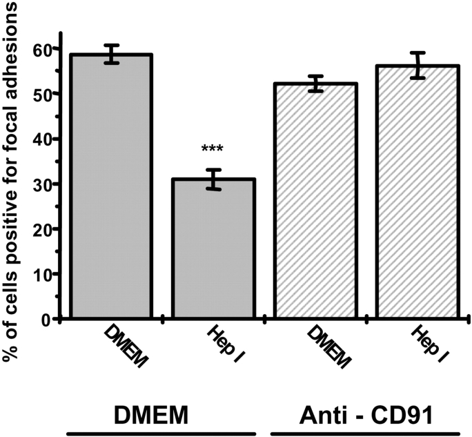
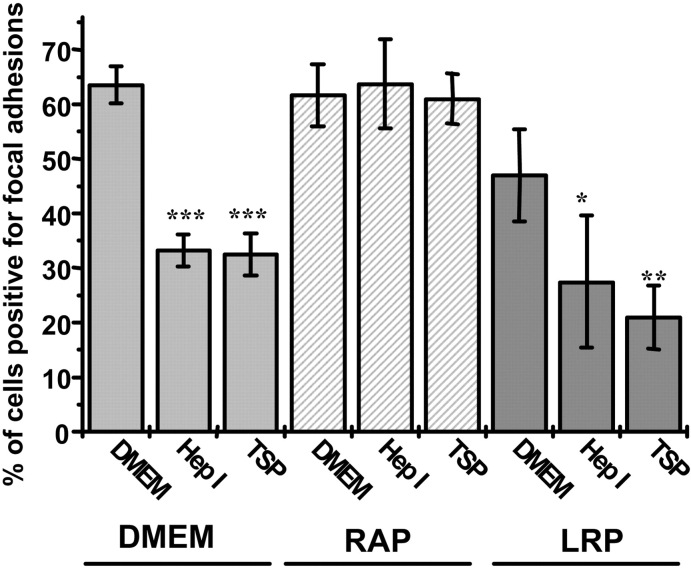
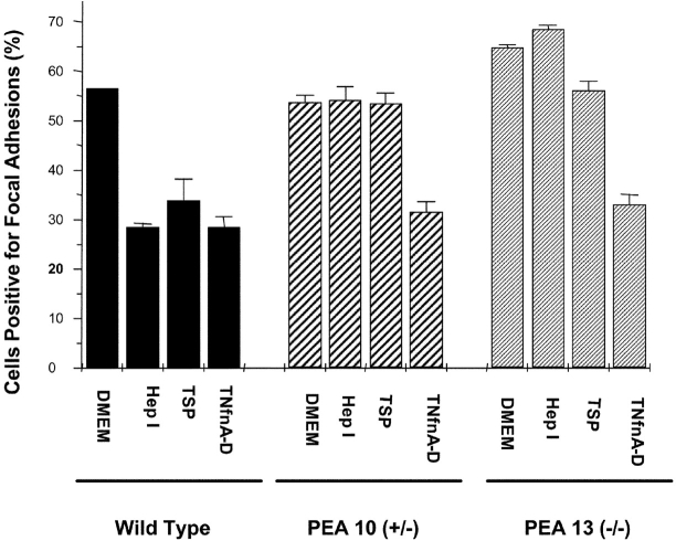
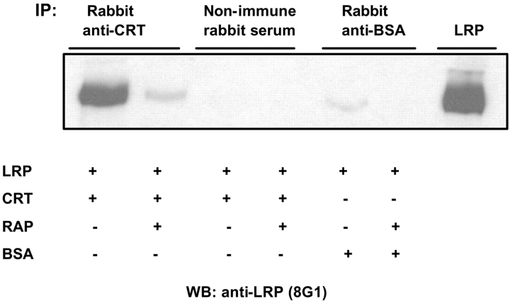
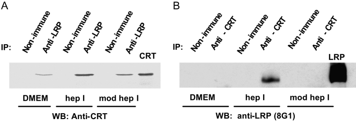
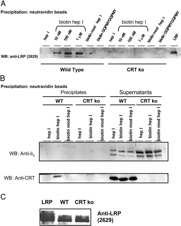
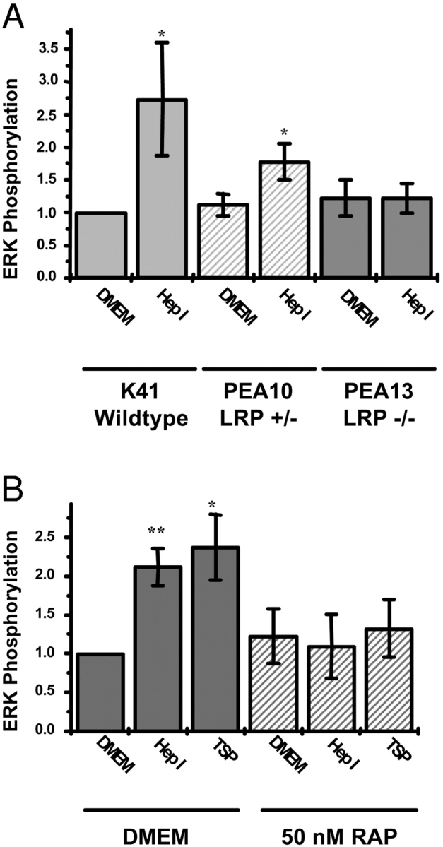
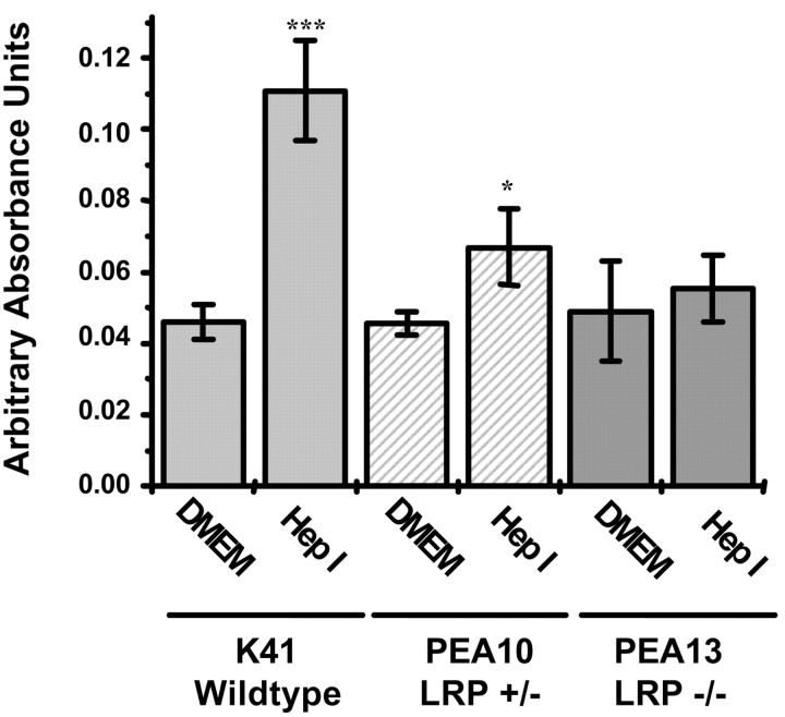
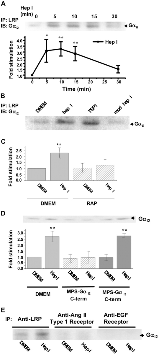
Thrombospondin (TSP) signals focal adhesion disassembly (the intermediate adhesive state) through interactions with cell surface calreticulin (CRT). TSP or a peptide (hep I) of the active site induces focal adhesion disassembly through binding to CRT, which activates phosphoinositide 3-kinase (PI3K) and extracellular signal-related kinase (ERK) through Galphai2 proteins. Because CRT is not a transmembrane protein, it is likely that CRT signals as part of a coreceptor complex. We now show that low density lipoprotein receptor-related protein (LRP) mediates focal adhesion disassembly initiated by TSP binding to CRT. LRP antagonists (antibodies, receptor-associated protein) block hep I/TSP-induced focal adhesion disassembly. LRP is necessary for TSP/hep I signaling because TSP/hep I is unable to stimulate focal adhesion disassembly or ERK or PI3K signaling in fibroblasts deficient in LRP. LRP is important in TSP-CRT signaling, as shown by the ability of hep I to stimulate association of Galphai2 with LRP. The isolated proteins LRP and CRT interact, and LRP and CRT are associated with hep I in molecular complexes extracted from cells. These data establish a mechanism of cell surface CRT signaling through its coreceptor, LRP, and suggest a novel function for LRP in regulating cell adhesion.


Figures

Figure 1.
Figure 1.
Anti-LRP antibody inhibits hep I–induced focal adhesion disassembly. BAE cells grown on coverslips were incubated with rabbit anti-LRP antibody (anti-CD91) for 30 min, washed, and then incubated with 1 μM hep I or DMEM (control) for 30 min. Cells were examined by interference reflection microscopy and assayed for the percentage of cells positive for focal adhesions ± SD (n = 3). A minimum of 300 cells per condition were evaluated. ***, P < 0.001 vs. DMEM.
Figure 2.
Figure 2.
RAP (but not LRP) inhibits TSP/hep I–induced focal adhesion disassembly. BAE cells grown on coverslips were incubated for 30 min with 2 μM RAP or DMEM (control) before addition of 100 nM hep I or 68 nM TSP for 30 min. In addition, LRP at 10-fold molar excess to 100 nM hep I or 340 nM TSP was incubated with 10 nM hep I or 34 nM TSP for 30 min before addition to cells for 30 min. Cells were fixed and examined for the number of cells positive for focal adhesions by interference reflection microscopy. Results are the mean ± SD (n = 3). *, P < 0.05; **, P < 0.01; ***, P < 0.001 vs. DMEM.
Figure 3.
Figure 3.
hep I does not induce focal adhesion disassembly. LRP-deficient wild-type (CRL-2214), PEA 10 (LRP +/−), and PEA 13 (LRP −/−) cells grown on coverslips were incubated with 1 μM hep I, 68 nM TSP, 30 μg/ml TN-C fnIIIA-D, or DMEM (untreated) for 30 min at 37°C. Cells were fixed and examined for focal adhesions by interference reflection microscopy in at least 250 cells/coverslip. Results are the mean percentage of cells positive for focal adhesions ± SD (n = 3).
Figure 4.
Figure 4.
Recombinant CRT interacts with purified LRP. 20 nM CRT and 10 nM LRP were incubated together in the presence or absence of 10 nM RAP, and then complexes were immunoprecipitated with anti-CRT antiserum or nonimmune rabbit serum. Alternately, LRP was incubated with BSA, and LRP bound to BSA was immunoprecipitated with anti-BSA antibody. LRP bound to CRT or BSA was detected after electrophoresis and transfer to nitrocellulose membranes with anti-LRP antibody (8G1).
Figure 5.
Figure 5.
LRP and CRT form complexes in BAE cells. (A) BAE cells were treated with DMEM, 1 μM hep I, or 1 μM modified hep I for 10 min, and N-octylglucopyranoside membrane extracts were immunoprecipitated with mouse anti-LRP antibody (5A6) or nonimmune IgG bound to GammaBind G Sepharose beads. Bound complexes were resolved by SDS-PAGE under nonreducing conditions, and CRT was detected by immunoblotting with goat–anti-CRT IgG. (B) BAE cells were incubated with either DMEM, 1 μM hep I, or 1 μM modified hep I for 10 min, and detergent extracts of membranes were immunoprecipitated with anti-CRT antiserum or nonimmune rabbit serum; bound LRP was detected by immunoblotting with mouse anti-LRP antibody (8G1).
Figure 6.
Figure 6.
LRP forms molecular complexes with biotin-tagged hep I peptide in wild-type (but not CRT-null) cell extracts. (A) Wild-type and CRT-null MEFs were incubated for 10 min with 1 μM untagged hep I peptide, 10 nM–1 μM biotin-tagged hep I, 1 μM biotin-tagged modified hep I, or 1 μM biotin-tagged CRT binding peptide. Detergent extracts of cells were coprecipitated with neutravidin beads to precipitate proteins associated with biotin peptides. LRP coprecipitating with the biotin–hep I complexes was detected by immunoblot with rabbit anti-LRP antibody (R2629). LRP was detected in the neutravidin– biotin precipitates from wild-type (but not from CRT-null) MEFs. LRP was not detected in samples treated with nontagged hep I, biotin modified hep I, or the biotin CRT–binding peptide. (B) Wild-type and CRT knockout MEFs were treated with 1 μM untagged hep I, biotin-tagged hep I, or biotin-tagged modified hep I, and associated proteins were precipitated with neutravidin. Precipitated proteins and proteins in the supernatant were assessed by immunoblotting for β3 integrin and then for CRT in the same membrane. (C) LRP levels were assessed by immunoblot in equivalent amounts of cell protein (50 μg) from wild-type and CRT-null MEFs.
Figure 7.
Figure 7.
TSP/hep I requires LRP to activate ERK. (A) Wild-type (K41), PEA10 (LRP+/−), and PEA13 (LRP−/−) MEFs were grown to near confluence in 6-well plates in 1% FBS overnight. Cells were incubated in serum-free DMEM cells (4 h) and then treated for 10 min with either 1 μM DMEM or 1 μM hep I. Cells lysates were analyzed for total and phospho-ERK by immunoblotting. Levels of phospho-ERK were determined through densitometry and normalized to total ERK levels. Results are the mean arbitrary absorbance units ± SD (n = 3–4). *, P < 0.05 vs. DMEM. (B) K41 MEFs were treated as in A, except that some cultures were treated with 50 nM RAP for the last hour of serum starvation. Cell lysates were probed for phosphoERK and total ERK as in A. Results are the mean arbitrary absorbance units ± SD (n = 3–4). *, P < 0.05; **, P < 0.01 vs. DMEM.
Figure 8.
Figure 8.
Hep I–mediated PI3K activation requires LRP. Wild-type K 41 MEFs, PEA10, and PEA13 cells were grown to near confluence, serum deprived overnight in 0.2% FBS, and treated with either 1 μM DMEM or 1 μM hep I for 30 min. Cells were lysed and immunoprecipitated with anti-p85 PI3K antibodies. Immunoprecipitates then underwent an in vitro lipid kinase assay by successive incubations with phosphatidylinositol-4,5-bisphosphate (PIP2) and [32P]ATP. Phosphorylated lipids were separated by TLC, detected by autoradiography, and analyzed using densitometry. Results are the mean arbitrary absorbance units ± SD (n = 3–5). *, P < 0.05; ***, P < 0.001 vs. DMEM.
Figure 9.
Figure 9.
Hep I stimulates association of LRP with the Gαi2 subunit, and association is blocked by RAP and by peptides corresponding to the COOH terminus of Gαi2. (A) BAE cells were grown to near confluence and serum deprived overnight. Cells were then treated with 1 μM hep I for 0, 5, 10, 15, or 30 min and lysed. Cell lysates were immunoprecipitated with monoclonal anti-LRP antibody (8G1), separated by SDS-PAGE, and immunoblotted with mouse anti-Gαi2 antibodies. A representative immunoblot for LRP-associated Gαi2 is shown. Bands were analyzed using One-Dscan software (Scanalytics), and the fold change in LRP-Gαi2 association as compared with untreated conditions was determined. Equal loading of sample protein was assessed by protein staining with Ponceau S. *, P < 0.05; **, P < 0.01 (n = 3–5). (B) BAE cells were grown as in A, treated for 10 min with either DMEM, 100 nM hep I, 78 nM TSP, or 100 nM modified hep I and assayed for LRP-Gαi2 association as described in A. A representative immunoblot for LRP-associated Gαi2 is shown (n = 3). (C) BAE cells were grown as in A and pretreated for 1 h in DMEM with or without 50 nM RAP and then treated with either DMEM or 1 μM hep I for 10 min and assayed for LRP-Gαi2 association as described in A. **, P < 0.01 (n = 4). (D) BAE cells were pretreated for 1 h in DMEM with or without 1 μM membrane-permeable sequence (MPS)-Gαi2 or MPS-Gαi3 peptides. Cells were then treated with either DMEM or 1 μM hep I for 10 min and lysed. Cells were then treated with either DMEM or 1 μM hep I for 10 min and assayed for LRP-Gαi2 association as described in A. **, P < 0.01 (n = 3–5). (E) BAE cells were treated with either DMEM or 1 μM hep I for 10 min and lysed. Cell lysates were immunoprecipitated with anti-LRP antibody (8G1), anti-angiotensin II type 1 receptor antibody, or anti-EGF receptor antibody, separated by SDS-PAGE, and immunoblotted with mouse anti-Gαi2 antibodies. Results are representative of at least three experiments.

References

    1. Arosa, F.A., O. de Jesus, G. Porto, A.M. Carmo, and M. de Sousa. 1999. Calreticulin is expressed on the cell surface of activated human peripheral blood T lymphocytes in association with major histocompatibility complex class I molecules. J. Biol. Chem. 274:16917–16922. - PubMed
    1. Ashcom, J.D., S.E. Tiller, K. Dickerson, J.L. Cravens, W.S. Argraves, and D.K. Strickland. 1990. The human alpha 2-macroglobulin receptor: identification of a 420-kD cell surface glycoprotein specific for the activated conformation of alpha 2-macroglobulin. J. Cell Biol. 110:1041–1048. - PMC - PubMed
    1. Aukhil, I., P. Joshi, Y. Yan, and H.P. Erickson. 1993. Cell- and heparin-binding domains of the hexabrachion arm identified by tenascin expression proteins. J. Biol. Chem. 268:2542–2553. - PubMed
    1. Baksh, S., and M. Michalak. 1991. Expression of calreticulin in Escherichia coli and identification of its Ca2+ binding domains. J. Biol. Chem. 266:21458–21465. - PubMed
    1. Barnes, H., B. Larsen, M. Tyers, and P. van Der Geer. 2001. Tyrosine-phosphorylated low density lipoprotein receptor-related protein 1 (LRP1) associates with the adapter protein SHC in SRC-transformed cells. J. Biol. Chem. 276:19119–19125. - PubMed

Publication types

MeSH terms

Substances