. 2003 Jul 8;100(14):8103-8.
doi: 10.1073/pnas.1032954100. Epub 2003 Jun 23.

Directed nucleation assembly of DNA tile complexes for barcode-patterned lattices

Affiliations

Directed nucleation assembly of DNA tile complexes for barcode-patterned lattices

Hao Yan et al. Proc Natl Acad Sci U S A. .

Abstract

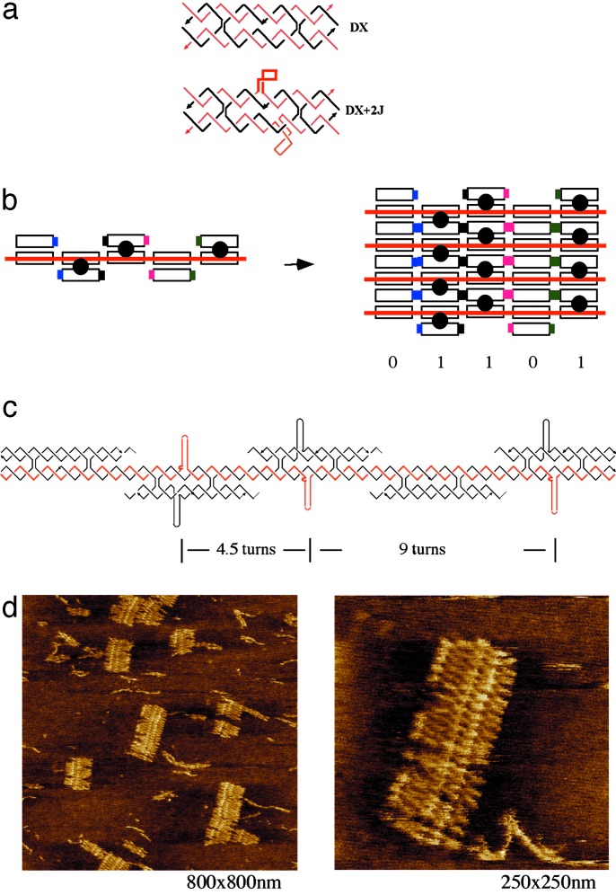
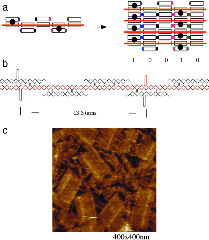
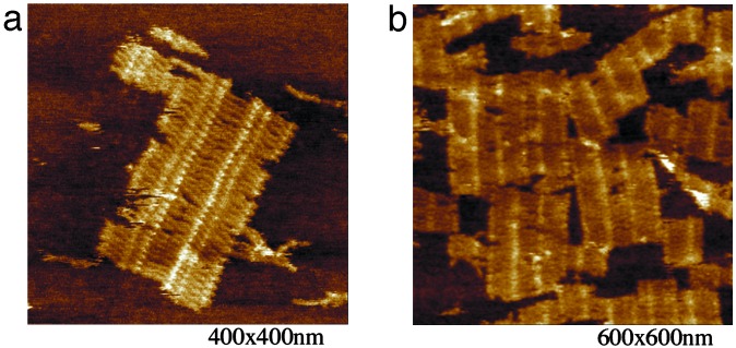
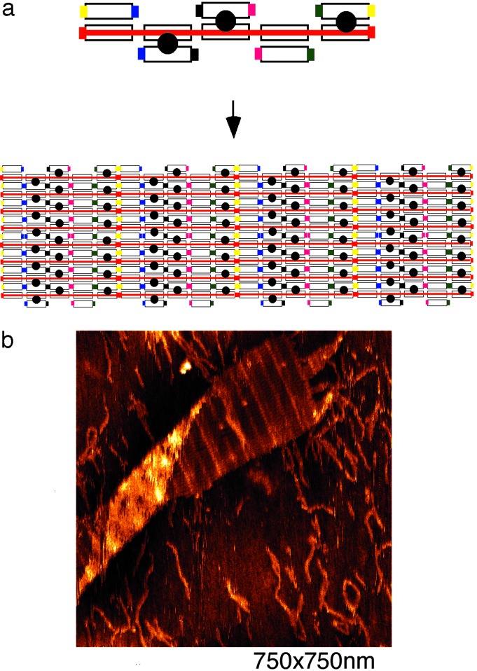
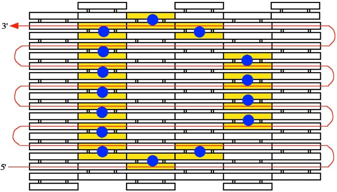
The programmed self-assembly of patterned aperiodic molecular structures is a major challenge in nanotechnology and has numerous potential applications for nanofabrication of complex structures and useful devices. Here we report the construction of an aperiodic patterned DNA lattice (barcode lattice) by a self-assembly process of directed nucleation of DNA tiles around a scaffold DNA strand. The input DNA scaffold strand, constructed by ligation of shorter synthetic oligonucleotides, provides layers of the DNA lattice with barcode patterning information represented by the presence or absence of DNA hairpin loops protruding out of the lattice plane. Self-assembly of multiple DNA tiles around the scaffold strand was shown to result in a patterned lattice containing barcode information of 01101. We have also demonstrated the reprogramming of the system to another patterning. An inverted barcode pattern of 10010 was achieved by modifying the scaffold strands and one of the strands composing each tile. A ribbon lattice, consisting of repetitions of the barcode pattern with expected periodicity, was also constructed by the addition of sticky ends. The patterning of both classes of lattices was clearly observable via atomic force microscopy. These results represent a step toward implementation of a visual readout system capable of converting information encoded on a 1D DNA strand into a 2D form readable by advanced microscopic techniques. A functioning visual output method would not only increase the readout speed of DNA-based computers, but may also find use in other sequence identification techniques such as mutation or allele mapping.


Figures

Fig. 1.
Fig. 1.
Self-assembly of 01101 barcode lattice around scaffold DNA strand. (a) (Upper) DAE tile, one type of antiparallel DNA DX tile. The tile drawing shows the five strands (three black and two red). The two red strands are continuous strands going through the tile in opposite directions (arrowheads mark 3′ ends). There are two crossover points connecting the two domains. There are two helical turns between the two crossover points. (Lower) DAE + 2J tile. This tile type has two hairpin loops protruding out of the central helix region of the DAE complex; one loop (thick line) is coming out of the plane and the other (thinner line) into the plane. The hairpin loops serve as topographic markers in AFM imaging of the lattices. (b) Schematic of self-assembly of barcode lattice layers based on DAE tiles around a scaffold strand. (Left) A five-tile crenellated horizontal layer is shown with an input scaffold strand running through the layer (red). The scaffold strand is required for the tiles to assemble. (Right) A lattice of four layers is illustrated (note that sticky ends are still available on the upper and lower layers for addition of more layers). The sticky ends are represented by different colored pads matching one other. The barcode information (01101) is represented by either the presence (designated 1) or the absence (designated 0) of a stem loop (shown as a black circle) protruding out of the tile plane. (c) Strand structure of one barcode layer. This layer represents barcode information of 01101. The red strand is the scaffold strand required for the tile assembly. The distance between adjacent hairpin loops is indicated by the number of helical turns. (d) AFM visualization of DNA barcode lattice (01101). The scale of each image is indicated in its lower right corner. Up to 24 layers of DNA have been self-assembled; the desired stripe pattern is clearly visible. Each layer contains five DX tiles and is ≈75 nm wide. The distance between the two closer adjacent stripes is ≈16 nm. The distance between the two further adjacent stripes is ≈31 nm. See Fig. 9, which is published as supporting information on the PNAS web site, for a large-area scan AFM image.
Fig. 2.
Fig. 2.
Inverse pattern barcode lattice (10010). (a) Schematic drawing of the barcode lattice representing the bit sequence 10010, which is the inverse of the first barcode of 01101. A single layer is shown (Left), and a four-layer lattice fragment is given (Right). (b) Strand structure of one barcode layer that represents barcode information of 01101. The red strand is the scaffold strand required for the tile assembly. The distance between adjacent hairpins is indicated in helical turns. (c) An AFM image at scale of 400 × 400 nm. Each layer contains five DX tiles, ≈75 nm wide, and the distance between the two stripes (designated 1) is ≈45 nm, as expected. See Fig. 9 for a large-area scan AFM image.
Fig. 3.
Fig. 3.
AFM images showing blunt-end helix stacking of tile assemblies. The scale is indicated below each image. (a) AFM image showing the alignment of two pieces of the first barcode lattice (01101) oriented in opposite directions. (b) AFM image showing the alignment of fragments of the second barcode lattice (10010) to form larger pieces of lattice.
Fig. 4.
Fig. 4.
Ribbon lattice formed from repeating DNA barcode units. (a) Schematic drawing showing the concatenation of the single-unit aperiodic barcode units into a repeating pattern. Two more pairs of sticky ends were included on the ends of the tile layer (compare with Fig. 1b). The sticky ends are represented by colored pads (yellow and red). (b) An AFM image showing the ribbon lattice displaying a repeating pattern of the barcode information. Distances between stripes are as expected.
Fig. 5.
Fig. 5.
Schematic drawing of a proposed 2D pattern using a directed nucleation assembly technique. In red is an input single-stranded DNA scaffold strand that encodes a 2D pattern (each odd row traversing from left to right, and each even row traversing right to left). Specific DNA tiles self-assemble around each segment of this input strand. The tiles then (or perhaps concurrently) self-assemble into a 2D tiling lattice with the predetermined pattern.

References

    1. Bernholc, J., Brenner, D., Nardelli, M. B., Meunier, V. & Roland, C. (2002) Annu. Rev. Mater. Res. 32 347-375.
    1. Joachim, C., Gimzewski, J. K. & Aviram, A. (2000) Nature 408 541-548. - PubMed
    1. Seeman, N. C. (2003) Nature 421 427-431. - PubMed
    1. LaBean, T. H. (2003) in Computational Biology and Genome Informatics, eds. Wang, J. T. L., Wu, C. H. & Wang, P. P. (World Scientific, River Edge, NJ), pp. 35-38.
    1. Reif, J. H. (2002) Comput. Sci. Eng. Mag.: Special Issue on Bio-Computation 4 32-41.

Publication types