. 2003 Jul 8;100(14):8583-8.
doi: 10.1073/pnas.1336802100. Epub 2003 Jun 23.

Phot1 and phot2 mediate blue light-induced transient increases in cytosolic Ca2+ differently in Arabidopsis leaves

Affiliations

Phot1 and phot2 mediate blue light-induced transient increases in cytosolic Ca2+ differently in Arabidopsis leaves

Akiko Harada et al. Proc Natl Acad Sci U S A. .

Abstract

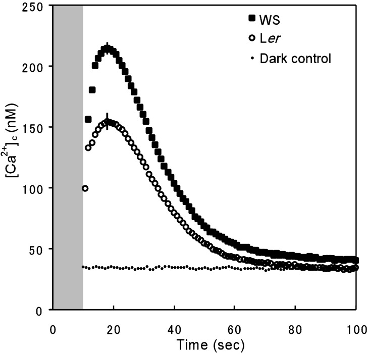
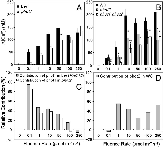
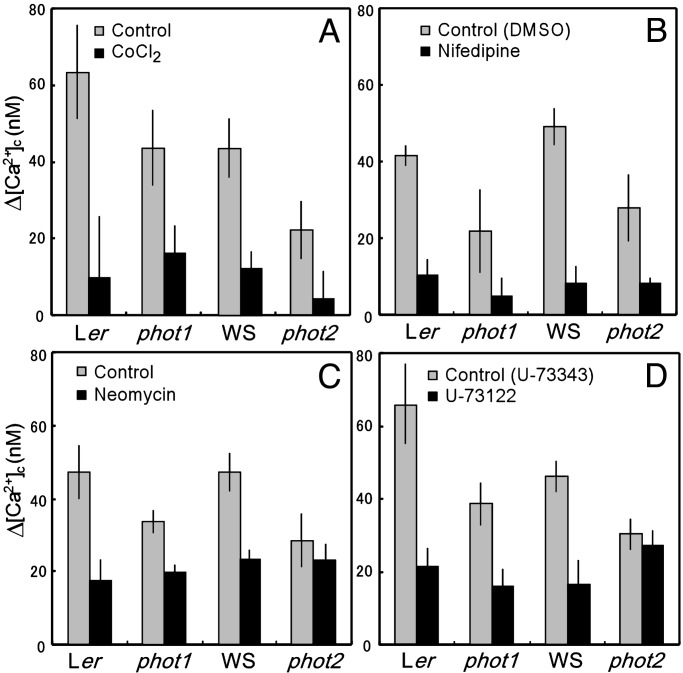
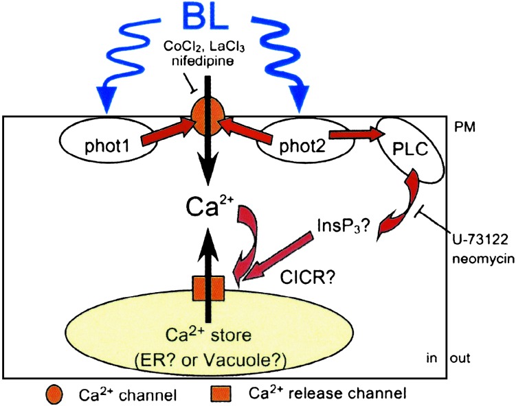
Phototropins (phot1 and phot2) are blue light (BL) receptors that mediate phototropism, chloroplast movements, and stomatal opening in Arabidopsis thaliana. Physiological studies have suggested that Ca2+ in the cytoplasm plays a pivotal role in these BL-induced responses. A phot1-mediated increase in cytosolic Ca2+ was reported in deetiolated seedlings of A. thaliana; however, the contribution of phot2 remains unknown. We examined a BL-induced transient increase in cytosolic free Ca2+ in leaves of transgenic A. thaliana of WT plants, phot1 and phot2 mutants, and phot1 phot2 double mutants expressing the Ca2+-sensitive luminescent protein aequorin. phot1 and phot2 had different photosensitivities: phot1 increased cytosolic Ca2+ at lower fluence rates (0.1-50 micromol x m-2 x s-1) and phot2 increased it at higher fluence rates (1-250 micromol x m-2 x s-1). By using Ca2+ channel blockers, Ca2+ chelating agents, and inhibitors of phospholipase C, we further demonstrated that both phot1 and phot2 could induce Ca2+ influx from the apoplast through the Ca2+ channel in the plasma membrane, whereas phot2 alone induced phospholipase C-mediated phosphoinositide signaling, which might result in Ca2+ release from internal Ca2+ stores. These results suggest that phot1 and phot2 mediate the BL-induced increase in cytosolic free Ca2+ differently.


Figures

Fig. 1.
Fig. 1.
BL-induced changes in [Ca2+]c. The averaged increases in [Ca2+]c were obtained from 10 independent experiments in leaves from Ler (circles) and WS (squares) plants. Leaves were illuminated for 10 s (gray zone) with 100 μmol·m-2·s-1 of BL. The vertical bars indicate the standard error. Dots represent dark control ([Ca2+]c in the same plants before illumination in Ler plants).
Fig. 2.
Fig. 2.
BL-induced increase in [Ca2+]c (Δ[Ca2+]c, the difference between [Ca2+]c in the dark before illumination and the BL-induced peak in [Ca2+]c)in leaves of WT plants, phot1 and phot2 mutants, and phot1 phot2 double mutants and relative contributions of phot1 and phot2 to the BL responses. (A and B) Δ[Ca2+]c in leaves of Ler plants and a phot1 mutant (A) and those in leaves of WS plants and phot2 and phot1 phot2 mutants (B) at various fluence rates of BL. (C and D) Relative contributions of phot1 (C) and phot2 (D) to the BL-induced Δ[Ca2+]c. The contributions of phot1 in the presence and absence of phot2 were calculated as the percent difference in Δ[Ca2+]c between phot1 and Ler (i.e., the results in A) and between phot1 phot2 and phot2 (results in B), respectively. The contribution of phot2 was calculated as the percent difference in Δ[Ca2+]c between phot2 and WS. The vertical bars indicate the standard error of 5–20 independent experiments. * and **, significantly different from the data in the WT plant and the phot2 mutant, respectively (P < 0.05).
Fig. 3.
Fig. 3.
Effects of inhibitors on BL-induced Δ[Ca2+]c in the WT plants and phot1 and phot2 mutants. A leaf, treated with each inhibitor in the dark, was irradiated with 1 μmol·m-2·s-1 of BL. Effects of 5 mM CoCl2 (A), 100 μM nifedipine (B; 0.4% DMSO was used as the control), 10 μM neomycin (C), and 10 μM U-73122 (D; 10 μM U-73343, an inactive analogue, was used as the control) are shown. The vertical bars indicate the standard error of 3–18 independent experiments.
Fig. 4.
Fig. 4.
Immunodetection of HA-PHOT1 and -PHOT2 from inner membrane-rich (IM) and PM-rich (PM) fractions isolated from leaves of HA-PHOT1 and -PHOT2 transformants, respectively. The proteins (3 μg in each lane) were separated by SDS/PAGE. Epitope-tagged PHOT1 and PHOT2 were detected as slightly larger bands than predicted molecular masses of phot1 (112 kDa) and phot2 (102 kDa), respectively.
Fig. 5.
Fig. 5.
A schematic model of the signal pathway of phot1- and phot2-dependent increase in [Ca2+]c in leaves of A. thaliana. phot1 and phot2 in the PM could mediate Ca2+ influx through a Ca2+ channel in the PM. phot2 alone induces CICR from internal Ca2+ stores through PLC-mediated signaling. In WT leaves, phot1 possibly increases the phot2-dependent PLC pathway and suppresses phot2-dependent Ca2+ influx from the apoplast. We describe the possibility in Discussion. See text for details.

Similar articles

Cited by

References

    1. Lin, C. (2002) Plant Cell Suppl. 14 S207-S225. - PMC - PubMed
    1. Briggs, W. R. & Christie, J. M. (2002) Trends Plant Sci. 7 204-210. - PubMed
    1. Huala, E., Oeller, P. W., Liscum, E., Han, I. S., Larsen, E. & Briggs, W. R. (1997) Science 278 2120-2123. - PubMed
    1. Kagawa, T., Sakai, T., Suetsugu, N., Oikawa, K., Ishiguro, S., Kato, T., Tabata, S., Okada, K. & Wada M. (2001) Science 291 2138-2141. - PubMed
    1. Christie, J. M., Reymond, P., Powell, G. K., Bernasconi, P., Raibekas, A. A., Liscum, E. & Briggs, W. R. (1998) Science 282 1698-1701. - PubMed

MeSH terms