. 2003 Jul 8;100(14):8247-52.
doi: 10.1073/pnas.1431613100. Epub 2003 Jun 23.

Mdm2-dependent ubiquitination and degradation of the insulin-like growth factor 1 receptor

Affiliations

Mdm2-dependent ubiquitination and degradation of the insulin-like growth factor 1 receptor

Leonard Girnita et al. Proc Natl Acad Sci U S A. .

Abstract

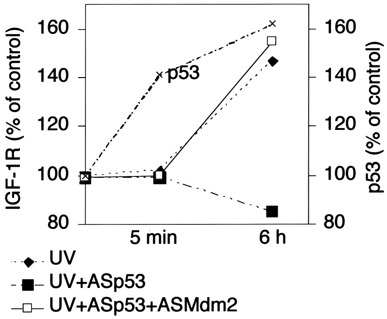
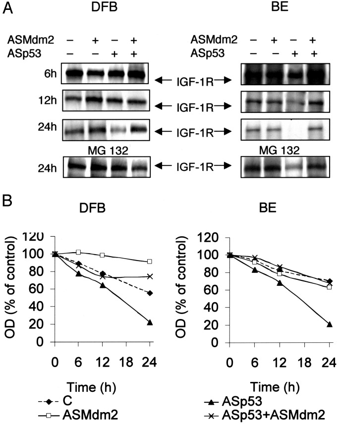
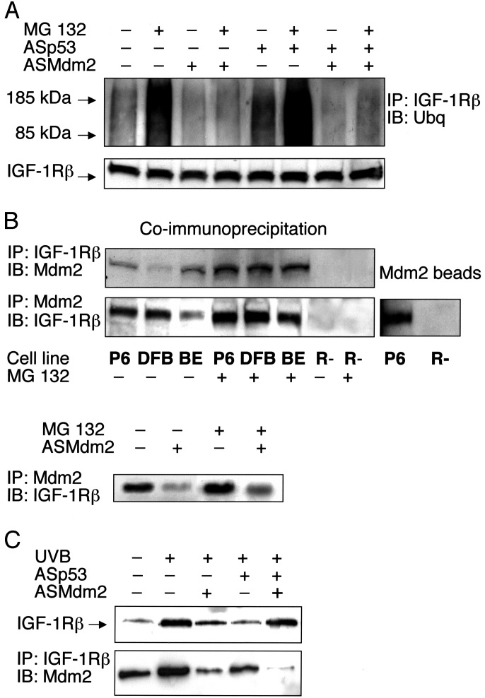
Recently, p53 was demonstrated to affect the expression of the insulin-like growth factor 1 receptor (IGF-1R), a receptor tyrosine kinase that plays a crucial role in growth and survival of cancer cells. However, the underlying mechanisms for interaction between p53 and IGF-1R are still not fully understood. One of the challenging questions remaining to be answered is why the wild-type p53, which per se represses the transcription of the IGF-1R gene, in overexpressed form is necessary for a high IGF-1R expression. In this study, we show that inhibition of p53 causes ubiquitination and down-regulation, through increased degradation, of the IGF-1R in human malignant melanoma cells. This effect, which was independent of the p53 status (i.e., wild type or mutated), was prevented if Mdm2 was coinhibited. Similar results were obtained in UV-irradiated human melanocytes (harboring wild-type p53), in which level of the IGF-1R increased after up-regulation of p53. Interestingly, the basal ubiquitination of the IGF-1R in untreated cells also depended on Mdm2. We could prove that Mdm2 physically associates with IGF-1R and that Mdm2 causes IGF-1R ubiquitination in an in vitro assay. Taken together our data provide evidence that Mdm2 serves as a ligase in ubiquitination of the IGF-1R and thereby causes its degradation by the proteasome system. Consequently, by sequestering Mdm2 in the cell nuclei, the level of p53 may indirectly influence the expression of IGF-1R. This role of Mdm2 and p53 represents an unexpected mechanism for the regulation of IGF-1R and cell growth.


Figures

Fig. 1.
Fig. 1.
Effect of p53 and Mdm2 inhibition on IGF-1R and cell growth on malignant melanoma cells. (A and B) The indicated cell lines either remained untreated (C) or were treated with Lipofectin (Lipofectin control, L), L + ASp53 (1.0 μM), L + ASMdm2 (2.0 μM), or L + ASp53 + ASMdm2 and with corresponding sense oligodeoxynucleotides (Sp53 and SMdm2) as indicated. After incubations for 24 h, the protein expression of p53, Mdm2, IGF-1R, and actin (loading control) was determined. Western blots for MEL-5 and MEL-28 are shown in A, and densitometry data (expressed as % of Lipofectin control) for all four cell lines are shown in B.(C) All four cell lines were treated as described in A but for 48 h. The amounts of surviving cells were determined by the XTT assay. The values are means and SDs of triplicates. It was confirmed in all experiments that Sp53 and SMdm2 were without effects (not shown in B and C). The experiments were repeated three to four times with similar results.
Fig. 2.
Fig. 2.
Effect of UV irradiation on IGF-1R expression of cultured melanocytes and effect of inhibition of p53 and Mdm2. Control cells or cells treated with ASp53 (1 μM) or ASMdm2 (1 μM) were exposed to 5 J/m2 UVB. Western blotting was performed to measure IGF-1R, and quantification was made by densitometry. The p53 levels (also assayed by Western blotting and densitometry) of irradiated control (not treated with AS) cells are indicated in the graph. For further details, see Materials and Methods. The experiment was repeated twice with similar results.
Fig. 3.
Fig. 3.
Effect of p53 and Mdm2 inhibition on IGF-1R degradation. (A) Wild-type (DFB) and p53 mutant (BE) cells were labeled with [35S]methionine (100 μCi/ml) for 24 h in methionine-free medium, and thereafter transferred to radioactive-free methionine-supplemented medium containing ASp53, ASMdm2, or both, or corresponding Ss for the indicated times. In separate dishes, cells incubated for 24 h were treated with MG 132 (50 μM) during the last 6 h (Bottom). IGF-1R β-subunit was immunoprecipitated and resolved by SDS/PAGE and finally visualized by autoradiography. It was confirmed that the sense oligodeoxynucleotides (Sp53 and SMdm2) were without effects (not shown). (B) Densitometry data are shown. The results are representative of three independent experiments.
Fig. 4.
Fig. 4.
Ubiquitination of IGF-1R and association with Mdm2. (A) DFB cells either remained untreated or were treated with ASp53, ASMdm2, or both as indicated for 12 h, after which the proteasome inhibitor MG 132 (50 μM) was added, or not, for an additional 6 h. IGF-1R was immunoprecipitated by a β-subunit antibody. Equal amounts of purified IGF-1R immunoprecipitates (to compensate for decrease in ASp53-treated cells), verified in the lower panel, were resolved by SDS/PAGE and finally detected by Western blotting using an antibody to ubiquitin. (B Upper) Total proteins isolated from the indicated cell lines were immunoprecipitated with an IGF-1R β-subunit antibody and immunoblotted by an Mdm2 antibody, or vice versa. (B Lower Left) BE cells were pretreated with ASMdm2 for 12 h, and the proteasome inhibitor MG 132 was added, or not, for an additional 6 h before coimmunoprecipitation. (B Right) Total protein lysates from P6 and R- cells were mixed with Sepharose beads of recombinant Mdm2 (see Materials and Methods). The Mdm2 beads were analyzed by Western blotting using an IGF-1R β-subunit antibody. (C Upper) UV-irradiated human melanocytes were treated as indicated for 6 h. Thereafter, analysis of IGF-1R was performed as described for the experiments on the melanoma cell lines. (C Lower) UV-irradiated melanocytes were treated as indicated for 6 h. Immunoprecipitation using an IGF-1R β-subunit antibody and immunoblotting with an Mdm2 antibody was then done. The experiments were repeated two to four times with similar results.
Fig. 5.
Fig. 5.
Mdm2-dependent ubiquitination of IGF-1R. (A Left) P6 cells either remained untreated or were treated with ASMdm2 for 18 h, with or without the proteasome inhibitor during the last 6 h. Equal amounts of purified immunoprecipitated IGF-1R β-subunit were analyzed by Western blotting using a ubiquitin antibody. The OD values for ubiquitinated IGF-1R are shown in A Lower.(A Right) UV-irradiated melanocytes either remained untreated or were treated with ASMdm2 as indicated for 6 h, whereupon immunoprecipitation and Western blot using ubiquitin antibodies were carried out. (B) In vitro ubiquitination of IGF-1R. Sepharose beads of IGF-1R, isolated from P6, were mixed with ubiquitin reagents with or without GST-Mdm2 as described in Materials and Methods. Equal amounts of purified IGF-1R immunoprecipitates (verified in Lower) were resolved by SDS/PAGE and finally detected by Western blotting using an antibody to His-tagged ubiquitin. The results are representative of three independent experiments.

References

    1. Baserga, R. (1995) Cancer Res. 55 249-252. - PubMed
    1. Baserga, R., Resnicoff, M. & Dews, M. (1997) Endocrine 7 99-102. - PubMed
    1. Werner, H. & Le Roith, D. (1997) Crit. Rev. Oncog. 8 71-92. - PubMed
    1. Ullrich, A., Gray, A., Tam, A. W., Yang-Feng, T., Tsubokawa, M., Collins, C., Henzel, W., Le Bon, T., Kathuria, S., Chen, E., et al. (1986) EMBO J. 5 2503-2512. - PMC - PubMed
    1. Buckbinder, L., Talbott, R., Velasco-Miguel, S., Takenaka, I., Faha, B., Seizinger, B. R. & Kley, N. (1995) Nature 377 646-649. - PubMed

Publication types

MeSH terms