. 2003;5(4):R186-92.
doi: 10.1186/ar762. Epub 2003 Apr 28.

Gene expression induced by interleukin-17 in fibroblast-like synoviocytes of patients with rheumatoid arthritis: upregulation of hyaluronan-binding protein TSG-6

Affiliations

Gene expression induced by interleukin-17 in fibroblast-like synoviocytes of patients with rheumatoid arthritis: upregulation of hyaluronan-binding protein TSG-6

A Kehlen et al. Arthritis Res Ther. 2003.

Abstract

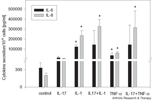
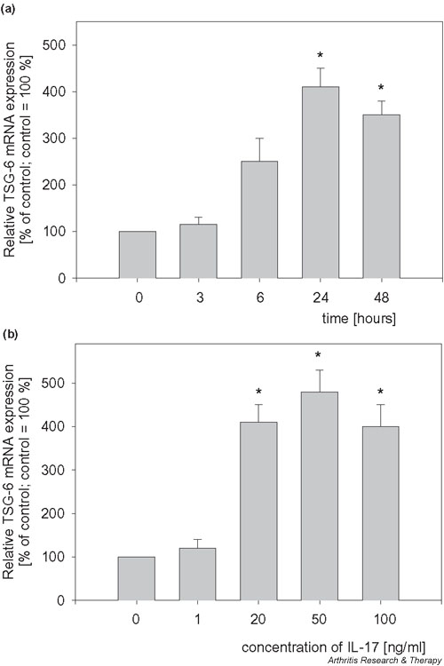
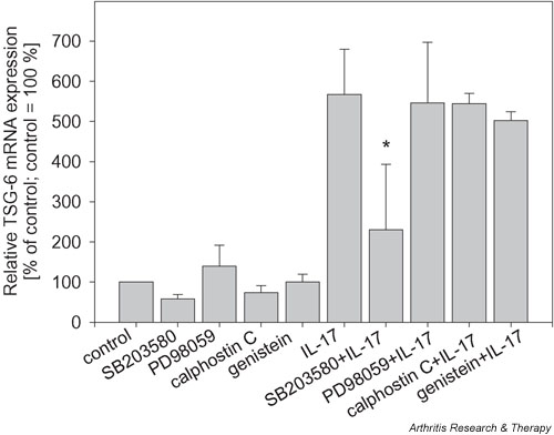
Interleukin-17 (IL-17) has been characterized as a proinflammatory cytokine produced by CD4+ CD45RO+ memory T cells. Overproduction of IL-17 was detected in the synovium of patients with rheumatoid arthritis (RA) compared with patients with osteoarthritis. This study examines differentially expressed genes after the stimulation of fibroblast-like synoviocytes of RA patients by IL-17. Among these genes we identified the following: tumor necrosis factor-stimulated gene-6 (TSG-6), IL-6, IL-8, GRO-beta, and bone morphogenetic protein-6 with an expression 3.6-10.6-fold that in the unstimulated control. IL-17 augmented the expression of TSG-6, a hyaluronan-binding protein, in a time- and dose-dependent manner. IL-17 showed additive effects with IL-1beta and tumour necrosis factor-alpha on the expression of TSG-6, IL-6 and IL-8. The mitogen-activated protein kinase p38 seems to be necessary for the regulation of TSG-6 expression by IL-17, as shown by inhibition with SB203580. Our results support the hypothesis that IL-17 is important in the pathogenesis of RA, contributing to an unbalanced production of cytokines as well as participating in connective tissue remodeling.


Figures

Figure 1
Figure 1
IL-17-induced gene expression. Fibroblast-like synoviocytes were cultured for 24 hours in the presence of IL-17 (20 ng/ml). Results of quantitative RT–PCR. Six different patients were measured each in duplicate, apart from nine different patients for TSG-6 measurements. *P < 0.05 in comparison with the unstimulated control.
Figure 2
Figure 2
Combined effects of IL-17 (20 ng/ml) plus IL-1β (10 ng/ml) or IL-17 plus TNF-α (10 ng/ml) on the expression of IL-6 and IL-8. Fibroblast-like synoviocytes were cultured for 72 hours in the presence of cytokines, and the concentrations of IL-6 or IL-8 in supernatants were determined. Measurements were made on synoviocytes from four different patients. *P < 0.05 in comparison with the results of IL-17 stimulation.
Figure 3
Figure 3
IL-17 stimulates the expression of TSG-6 mRNA. (a) Time course of TSG-6 mRNA expression after stimulation with 20 ng/ml IL-17. (b) Dose-dependent stimulation of TSG-6 mRNA expression after stimulation with IL-17 for 24 hours. Results of quantitative RT–PCR are given as percentages of the basal control (culture without IL-17 set at 100%). Results are from four different patients, each measured in duplicate. *P < 0.05 in comparison with the unstimulated control.
Figure 4
Figure 4
Combined effects of IL-17 (20 or 50 ng/ml) plus IL-1β (10 ng/ml) or IL-17 (20 or 50 ng/ml) plus TNF-α (10 ng/ml) on the expression of TSG-6 mRNA. Results of quantitative RT–PCR are given as percentages of the basal control (culture without IL-17 set at 100%). Results are from six different patients, each measured in duplicate. *P < 0.05 in comparison with the results of IL-1 stimulation.
Figure 5
Figure 5
Combined effects of IL-17 (20 ng/ml) plus IL-1β (10 ng/ml) or IL-17 (20 ng/ml) plus TNF-α (10 ng/ml) on the expression of TSG-6 protein. Fibroblast-like synoviocytes (SFCs) were cultured for 48 hours with the cytokines indicated below. Cells were lysed, separated, blotted, and probed with a TSG-6 antibody as described in Materials and methods. Lanes 1–6, SFCs of patient 1; lanes 7–12, SFCs of patient 2. Lanes 1 and 7, control; lane 2, IL-17; lanes 3 and 8, IL-1β; lanes 4 and 9, IL-17 plus IL-1β; lanes 5 and 10, TNF-α; lanes 6 and 11, IL-17 plus TNF-α; lane 12, IL-17 plus anti-IL-17 antibody. The intensity was analyzed densitometrically and normalized to the intensity of the appropriate control.
Figure 6
Figure 6
Effects of protein kinase inhibitors on the expression of TSG-6 mRNA. Fibroblast-like synoviocytes were cultured for 24 hours with or without IL-17 (20 ng/ml) and the appropriate inhibitor. Total RNA (0.5 μg) was used for cDNA synthesis in a volume of 10 μl; 1.5 μl of the synthesized cDNA was used for real-time PCR as described. Results are given as a percentage of the basal control (culture without cytokine set at 100%). Results are from four different patients, each measured in duplicate. *P < 0.05 in comparison with the results of IL-17 stimulation.

References

    1. Firestein GS. Invasive fibroblast-like synoviocytes in rheumatoid arthritis. Passive responders or transformed aggressors? Arthritis Rheum. 1996;39:1781–1790. - PubMed
    1. Weyand CM, Goronzy JJ. The molecular basis of rheumatoid arthritis. J Mol Med. 1997;75:772–785. doi: 10.1007/s001090050167. - DOI - PubMed
    1. Breedveld FC. New insights in the pathogenesis of rheumatoid arthritis. J Rheumatol. 1998;53(suppl):S3–S7. - PubMed
    1. Feldmann M, Brennan FM, Maini RN. Role of cytokines in rheumatoid arthritis. Annu Rev Immunol. 1996;14:397–440. doi: 10.1146/annurev.immunol.14.1.397. - DOI - PubMed
    1. Feldmann M, Brennan FM, Maini RN. Rheumatoid arthritis. Cell. 1996;85:307–310. - PubMed

MeSH terms