. 2003 Jul 1;31(13):3386-92.
doi: 10.1093/nar/gkg578.

STING Millennium: A web-based suite of programs for comprehensive and simultaneous analysis of protein structure and sequence

Affiliations

STING Millennium: A web-based suite of programs for comprehensive and simultaneous analysis of protein structure and sequence

Goran Neshich et al. Nucleic Acids Res. .

Abstract

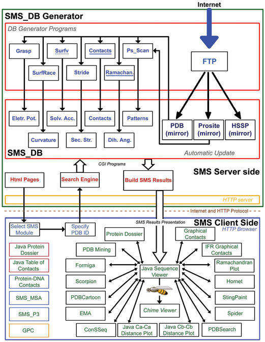
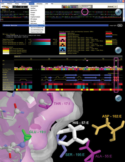
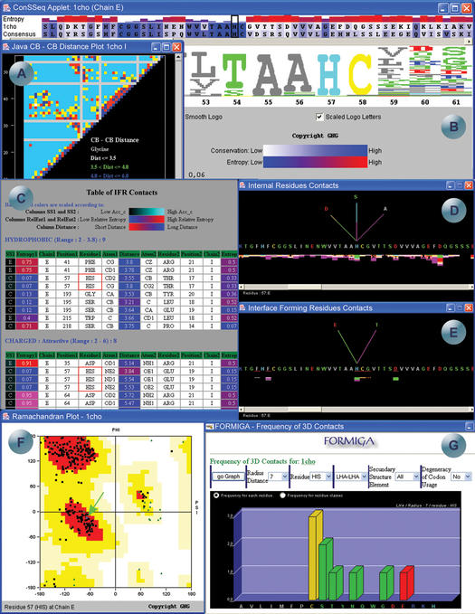
STING Millennium Suite (SMS) is a new web-based suite of programs and databases providing visualization and a complex analysis of molecular sequence and structure for the data deposited at the Protein Data Bank (PDB). SMS operates with a collection of both publicly available data (PDB, HSSP, Prosite) and its own data (contacts, interface contacts, surface accessibility). Biologists find SMS useful because it provides a variety of algorithms and validated data, wrapped-up in a user friendly web interface. Using SMS it is now possible to analyze sequence to structure relationships, the quality of the structure, nature and volume of atomic contacts of intra and inter chain type, relative conservation of amino acids at the specific sequence position based on multiple sequence alignment, indications of folding essential residue (FER) based on the relationship of the residue conservation to the intra-chain contacts and Calpha-Calpha and Cbeta-Cbeta distance geometry. Specific emphasis in SMS is given to interface forming residues (IFR)-amino acids that define the interactive portion of the protein surfaces. SMS may simultaneously display and analyze previously superimposed structures. PDB updates trigger SMS updates in a synchronized fashion. SMS is freely accessible for public data at http://www.cbi.cnptia.embrapa.br, http://mirrors.rcsb.org/SMS and http://trantor.bioc.columbia.edu/SMS.


Figures

Figure 1
Figure 1
Schematic diagram illustrating the architecture of SMS. SMS is organized in two logical layers: SMS server and SMS client. On the server side, illustrated inside the large green box, are carried out all updates of the public domain databases used by SMS and the subsequent calculation of macromolecular properties. The SMS client side, illustrated inside the large blue box, provides a user friendly graphical interface and communicates to SMS server. The SMS versions 2.2, 3.0, 3.1 and 4.0 components are listed within black, red, blue and yellow border boxes, respectively.
Figure 2
Figure 2
SMS example screens for the pdb file 1cho.pdb, highlighting the residue His57 in chain E. (A) Sequence frame, from where all the SMS modules and features can be accessed. The sequence is colored according to physico-chemical properties of the amino acids. The blue and red lines underneath the amino acid sequence represents the secondary structure elements (beta strands and helices respectively), according to the pdb file annotation. As the user scrolls the mouse over the sequence, information about the residues appears in the ‘residue info’ box. Two pull-down menus are also shown, demonstrating choice of SMS modules and rendering. (B) Protein Dossier module—the meaning of the colors are on the top part of the image. The internal and interface forming contacts are shown above the amino acid sequence cartoon, color coded according to the type of contact. The first row (red line) under the sequence highlights the residues at the interface. The next row (yellow line bellow the sequence stretch: VTAAHGC) indicates the PROSITE pattern PS00134. The following three rows indicate the secondary structure according to the annotations in PDB, DSSP and Stride, respectively. Another five rows are displayed color coded according to: temperature factor, relative entropy, accessibility in complex, accessibility in isolation and dihedral angles. His_57 and parameters belonging to this residue are placed within magenta quadrant. (C) 1cho structure representation rendered in the SMS structure window. The two surfaces represent the interface between the chain E (white) and chain I (pink). The figure was centered on the His57:E residue, painted in white. The other residues drawn in stick presentation are the residues that make contacts with the histidine, colored according to the contacts they make, corresponding to the first two lines of the Protein Dossier in (B). This particular image was obtained by combination of SMS actions, starting with the left mouse click on the Graphical IFR contacts java window, right above His_57 residue. Subsequently, some viewing arrangements are done in terms of removal of undesired information and then followed by surface building for the portion that makes part of the interface between the enzyme and inhibitor chains.
Figure 3
Figure 3
SMS example screens for the pdb file 1cho.pdb. (A) Java Cβ–Cβ distance plot, representing the distances between the beta carbons in chain I. (B) ConSSeq window where the amino acid sequence of 1cho, chain E is aligned with a consensus sequence as found in HSSP and, entropy histogram color and size coded with respect to the calculated degree of conservation. The size of sequence letters in the ‘logo’ follows the same degree of conservation, while color coding is the same as in SMS sequence frame. (C) Table of IFR contacts of the His_57, chain E amino acid. The table shows all the contacts in the protein, listing all the atoms along with its secondary structure, entropy, chain, residue number, residue name, atom name and distance in Å between two atoms in contact. The accessibility, entropy and distances of all atoms involved in the contacts are color coded and presented as background color. The accessibility is colored in the first column, ranging from black (low accessibility) to cyan (high accessibility), the entropy coloring scheme ranges from blue (low entropy) to red (high entropy) and the distances are colored from red (short) to blue (long). (D) Internal residue contacts formed by the His_57, chain E, showing all amino acids, belonging to the same chain E, that make contacts with it. The fan above the residue is color coded according to the type of contact the residue is involved with. (E) Interface forming residues contacts formed by the His_57, chain E. (F) Ramachandran plot of 1cho.pdb, highlighting the position of His_57, chain E in the diagram, and coloring plotted areas according to the allowed regions for the phi and psi angles. (G) Module FORMIGA showing the frequency of neighbors to the histidine residues (all His residues of E chain are considered and data demonstrated as a sum of calculated hits) found within the 7 Å radius sphere, centered at last heavy atom (LHA) of the histidine side chain.

References

    1. Berman H.H., Westbrook,J., Feng,Z., Gilliland,G., Bhat,T.N., Weissig,H., Shindyalov,I.N. and Bourne,P.E. (2000) The Protein Data Bank. Nucleic Acids Res., 28, 235–242. - PMC - PubMed
    1. Laskovski R.A., Hutchinson,E.G., Michie,A.D., Wallace,A.C., Jones,M.L. and Thornton,J.M. (1997) PDBsum: a web-based database of summaries and analyses of all PDB structures. Trends Biochem. Sci., 22, 488–490. - PubMed
    1. Hogue C.W. (1997) Cn3D: a new generation of three-dimensional molecular structure viewer. Trends Biochem. Sci., 22, 314–316. - PubMed
    1. Neshich G., Togawa,R.C. and Honig,B. (1998) Sequence To and withIN graphics PDB Viewer (STING-PDB viewer). PDB Quart. News., July (electronic edition), 6.
    1. Sander C. and Schneider,R. (1991) Database of homology-derived protein structures and the structural meaning of sequence alignment. Proteins, 9, 56–68. - PubMed

Publication types

Associated data