Review
. 2004 Jan 15;554(Pt 2):275-83.
doi: 10.1113/jphysiol.2003.047902. Epub 2003 Jun 24.

Physiological modulation of inactivation in L-type Ca2+ channels: one switch

Affiliations
Review

Physiological modulation of inactivation in L-type Ca2+ channels: one switch

Ian Findlay. J Physiol. .

Abstract

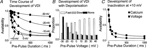
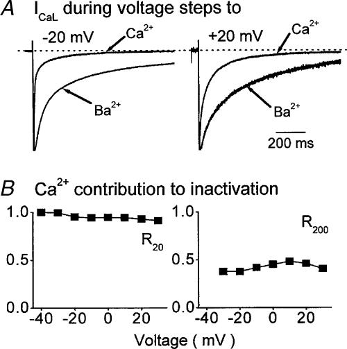
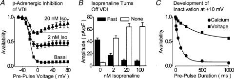
The relative contributions of voltage- and Ca(2+)-dependent mechanisms of inactivation to the decay of L-type Ca(2+) channel currents (I(CaL)) is an old story to which recent results have given an unexpected twist. In cardiac myocytes voltage-dependent inactivation (VDI) was thought to be slow and Ca(2+)-dependent inactivation (CDI) resulting from Ca(2+) influx and Ca(2+)-induced Ca(2+)-release (CICR) from the sarcoplasmic reticulum provided an automatic negative feedback mechanism to limit Ca(2+) entry and the contribution of I(CaL) to the cardiac action potential. Physiological modulation of I(CaL) by Beta-adrenergic and muscarinic agonists then involved essentially more or less of the same by enhancing or reducing Ca(2+) channel activity, Ca(2+) influx, sarcoplasmic reticulum load and thus CDI. Recent results on the other hand place VDI at the centre of the regulation of I(CaL). Under basal conditions it has been found that depolarization increases the probability that an ion channel will show rapid VDI. This is prevented by Beta-adrenergic stimulation. Evidence also suggests that a channel which shows rapid VDI inactivates before CDI can become effective. Therefore the contributions of VDI and CDI to the decay of I(CaL) are determined by the turning on, by depolarization, and the turning off, by phosphorylation, of the mechanism of rapid VDI. The physiological implications of these ideas are that under basal conditions the contribution of I(CaL) to the action potential will be determined largely by voltage and by Ca(2+) following Beta-adrenergic stimulation.


Figures

Figure 1
Figure 1. Inactivation of ICaL under basal conditions
A, normalized ICaL carried by Ca2+ and Ba2+ in one ventricular myocyte. At negative voltages (left) Ca2+ current inactivates more rapidly than Ba2+ current. At positive voltages (right) the initial decay of Ba2+ current is as fast as that of Ca2+ current, only later does Ca2+ current show more inactivation. B, the contribution of Ca2+ to the inactivation of ICaL evaluated by the ratio of decay recorded with Ca2+ relative to that recorded with Ba2+, 20 ms (R20, left) and 200 ms (R200, right) after activation. At both times the contribution of Ca2+ declines with depolarization though there is clearly more of an effect of Ca2+ at positive voltages after 200 ms than after 20 ms. The figure has been redrawn from Findlay (2002a).
Figure 2
Figure 2. The development of rapid VDI under basal conditions
A, the time course of the development of VDI was recorded in the absence of Ca2+, and in the absence of ion flux through the channels, between voltages which evoke minimal (–30 mV) and maximal (+10 mV) inactivation. The lines connecting data points represent the fitting of either a single (–30 and –20 mV) or a double (–10, 0 and +10 mV) exponential function to the data. B, the proportions of fast (open columns), slow (hatched columns) and no (filled columns) VDI which were recorded at different membrane potentials show that as the amount of current which showed fast VDI increased, that which showed no VDI declined. C, the relative contributions of Ca2+ and voltage to the decay of ICaL were assessed by measuring the time course of development of inactivation at +10 mV in the presence (circles) and the absence (squares) of extracellular Ca2+. The first recorded total inactivation, being the sum of VDI and CDI. The second recorded only VDI since no ion flux through ICaL occurred at this voltage under these conditions (see Findlay, 2002b for further details). It is clear that Ca2+ influx does little to increase the initial rapid phase of the development of inactivation. The results shown here were conducted in the presence of ryanodine. A and B have been redrawn from Findlay (2002d) and C from Findlay (2002b).
Figure 3
Figure 3. Inactivation of ICaL following β-adrenergic stimulation
A, normalized ICaL carried by Ca2+ and Ba2+ in one ventricular myocyte. At negative (left) and positive (right) voltages Ca2+ current inactivates more rapidly than Ba2+ current. B, the contribution of Ca2+ to the inactivation of ICaL 20 ms (R20, left) and 200 ms (R200, right) after activation is clearly sustained at all membrane voltages. The figure has been redrawn from Findlay (2002a).
Figure 4
Figure 4. Inhibition of rapid VDI by β-adrenergic stimulation
A, dose-dependent reduction of the availability–voltage relationship of ICaL by Isoprenaline (Iso). Experiments were conducted in the absence of extracellular Ca2+ with a double-pulse voltage-clamp protocol with 1000 ms duration prepulse voltage steps. B, the effects of isoprenaline upon the amount of ICaL which showed fast VDI (filled columns) and no inactivation (open columns). These experiments were conducted in the absence of extracellular Ca2+ and Findlay (2002c) should be consulted for further details. C, the relative contributions of Ca2+ and voltage to the decay of ICaL were assessed as described in Fig. 2C in the presence (circles) and the absence (squares) of extracellular Ca2+ in the presence of 100 nm isoprenaline. It is clear that Ca2+ influx dramatically increases the rate of development of inactivation, and comparison with Fig. 2C reveals that this is due to the suppression of fast VDI. These experiments were conducted in the presence of ryanodine. A and B have been redrawn from Findlay (2002c) and C from Findlay (2002b).

Similar articles

Cited by

References

    1. Adachi-Akahane S, Leeman L, Morad M. Cross-signalling between L-type Ca2+ channels and ryanodine receptors in rat ventricular myocytes. J General Physiol. 1996;108:435–454. - PMC - PubMed
    1. Adams B, Tanabe T. Structural regions of the cardiac Ca channel alpha subunit involved in Ca-dependent inactivation. J General Physiol. 1997;110:379–389. - PMC - PubMed
    1. Alseikhan BA, DeMaria CD, Colecraft HM, Yue DT. Engineered calmodulins reveal the unexpected eminence of Ca2+ channel inactivation in controlling heart excitation. Proc Natl Acad Sci USA. 2002;99:17185–17190. - PMC - PubMed
    1. Anderson ME. Ca2+-dependent regulation of cardiac L-type Ca2+ channels: is a unifying mechanism at hand. J Mol Cell Cardiol. 2001;33:639–650. - PubMed
    1. Antz C, Bauer T, Kalbacher H, Frank R, Covarrubias M, Kalbitzer HR, Ruppersberg JP, Baukrowitz T, Fakler B. Control of K+ channel gating by protein phosphorylation: structural switches of the inactivation gate. Nature Struc Biol. 1999;6:146–150. - PubMed

LinkOut - more resources