. 2003 Aug 15;551(Pt 1):5-12.
doi: 10.1113/jphysiol.2003.042002. Epub 2003 Jun 24.

Calmodulin kinase modulates Ca2+ release in mouse skeletal muscle

Affiliations

Calmodulin kinase modulates Ca2+ release in mouse skeletal muscle

Pasi Tavi et al. J Physiol. .

Abstract

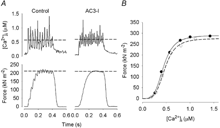
Activation of the contractile machinery in skeletal muscle is initiated by the action-potential-induced release of Ca2+ from the sarcoplasmic reticulum (SR). Several proteins involved in SR Ca2+ release are affected by calmodulin kinase II (CaMKII)-induced phosphorylation in vitro, but the effect in the intact cell remains uncertain and is the focus of the present study. CaMKII inhibitory peptide or inactive control peptide was injected into single isolated fast-twitch fibres of mouse flexor digitorum brevis muscles, and the effect on free myoplasmic [Ca2+] ([Ca2+]i) and force during different patterns of stimulation was measured. Injection of the inactive control peptide had no effect on any of the parameters measured. Conversely, injection of CaMKII inhibitory peptide decreased tetanic [Ca2+]i by ~25 %, but had no significant effect on the rate of SR Ca2+ uptake or the force-[Ca2+]i relationship. Repeated tetanic stimulation resulted in increased tetanic [Ca2+]i, and this increase was smaller after CaMKII inhibition. In conclusion, CaMKII-induced phosphorylation facilitates SR Ca2+ release in the basal state and during repeated contractions, providing a positive feedback between [Ca2+]i and SR Ca2+ release.


Figures

Figure 1
Figure 1. Inhibition of CaMKII decreases both tetanic [Ca2+]i and force
Representative original records of [Ca2+]i (upper row) and force (lower row) in 70-Hz contractions produced before and after injection of the active CaMKII inhibitory peptide (AC3-I; A) or the inactive control peptide (AC3-C; B). Myoplasmic concentrations of AC3-I and AC3-C were 11.5 and 21.0 μM, respectively.
Figure 2
Figure 2. Injection of CaMKII inhibitory peptide decreases tetanic [Ca2+]i
A, mean data (± S.E.M.) for [Ca2+]i during 70-Hz tetani vs. myoplasmic concentration of indo-1 obtained after injection of indo-1 only (▴; n = 13), the active inhibitory peptide (•; n = 7) and the inactive control peptide (○, n = 6). B, individual measurements of the relative decrease in tetanic [Ca2+]i after injection of the active peptide (•). The line shows a one-site saturation curve fitted to the data points. For comparison, mean data (± S.E.M.) from fibres injected with the inactive peptide are also shown.
Figure 3
Figure 3. Inhibition of CaMKII has no effect on the force-[Ca2+]i relationship
A, original records from one muscle fibre of [Ca2+]i (upper row) and force (lower row) obtained during a 30-Hz contraction before (left) and a 50-Hz contraction after (right) injection of the active inhibitory peptide (AC3-I). The dashed lines show mean [Ca2+]i and force, both of which were similar before and after injection. B, force-[Ca2+]i data points from the same fibre were obtained using 10- to 100-Hz tetani before (•) and after (○) injection of the peptide. A Hill equation (see Methods) was fitted to pre- (full line) and post-injection (dashed line) data points.
Figure 4
Figure 4. The force decrease after CaMKII inhibition is larger at low stimulation frequencies
Mean data (± S.E.M., n = 5) of relative [Ca2+]i (○) and force (•) at different tetanic stimulation frequencies, calculated as the ratio between the post- and pre-injection measurements of CaMKII inhibitory peptide. The dashed line indicates no change.
Figure 5
Figure 5. Inhibition of CaMKII decreases the increase in tetanic [Ca2+]i during a series of repeated tetani
Mean data (± S.E.M.) for tetanic [Ca2+]i during a series of 70-Hz tetanic contractions (350 ms duration, 2 s intervals) measured before (•) and after (○) injection of the active inhibitory peptide (A; n = 6) and the inactive control peptide (B; n = 4).
Figure 6
Figure 6. CaMKII phosphorylation increases during a series of repeated tetanic contractions
A, mean [Ca2+]i record from a series of 10 repeated 70-Hz tetani (350 ms duration, 2 s intervals) produced under control conditions. B, modelled relative phosphorylation of CaMKII. The [Ca2+]i record in A was used to drive the model.
Figure 7
Figure 7. CaMKII inhibition has no effect on the rate of decline of tetanic [Ca2+]i
Mean [Ca2+]i (± S.E.M.; n = 4-7) measured during relaxation following a single 70-Hz tetanus (A and B) and at the end of a series of ten 70-Hz tetani (C and D). Data were obtained before (•) and after (○) injection of the active (A and C) and inactive (B and D) peptide.

References

    1. Allen DG, Duty S, Westerblad H. Metabolic changes in muscle during exercise; their effect on muscle function. Proc Aust Physiol Soc. 1993;24:65–75.
    1. Allen DG, Westerblad H. Role of phosphate and calcium stores in muscle fatigue. J Physiol. 2001;536:657–665. - PMC - PubMed
    1. Anderson ME. Ca2+-dependent regulation of cardiac L-type Ca2+ channels: is a unifying mechanism at hand? J Mol Cell Cardiol. 2001;33:639–650. - PubMed
    1. Balog EM, Fruen BR, Kane PK, Louis CF. Mechanisms of Pi regulation of the skeletal muscle SR Ca2+ release channel. Am J Physiol. 2000;278:C601–611. - PubMed
    1. Bayer KU, Harbers K, Schulman H. α KAP is an anchoring protein for a novel CaM kinase II isoform in skeletal muscle. EMBO J. 1998;17:5598–5605. - PMC - PubMed

Publication types

MeSH terms

Substances

LinkOut - more resources