. 2003 Jul;112(1):42-9.
doi: 10.1172/JCI17856. Epub 2003 Jun 16.

Bone marrow stem cells contribute to repair of the ischemically injured renal tubule

Affiliations

Bone marrow stem cells contribute to repair of the ischemically injured renal tubule

Sujata Kale et al. J Clin Invest. 2003 Jul.

Abstract

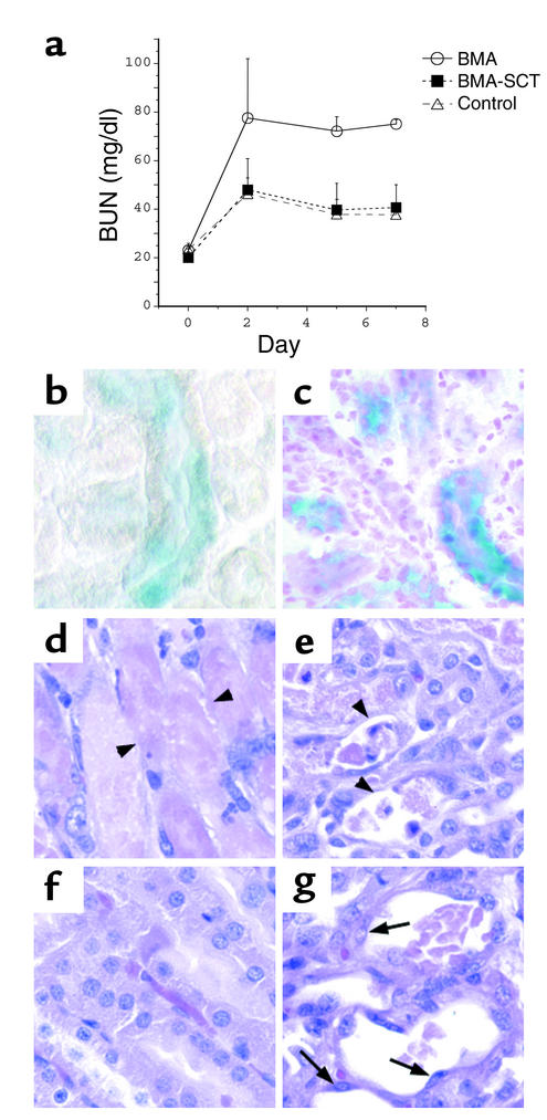
The paradigm for recovery of the renal tubule from acute tubular necrosis is that surviving cells from the areas bordering the injury must migrate into the regions of tubular denudation and proliferate to re-establish the normal tubular epithelium. However, therapies aimed at stimulating these events have failed to alter the course of acute renal failure in human trials. In the present study, we demonstrate that Lin-Sca-1+ cells from the adult mouse bone marrow are mobilized into the circulation by transient renal ischemia and home specifically to injured regions of the renal tubule. There they differentiate into renal tubular epithelial cells and appear to constitute the majority of the cells present in the previously necrotic tubules. Loss of stem cells following bone marrow ablation results in a greater rise in blood urea nitrogen after renal ischemia, while stem cell infusion after bone marrow ablation reverses this effect. Thus, therapies aimed at enhancing the mobilization, propagation, and/or delivery of bone marrow stem cells to the kidney hold potential as entirely new approaches for the treatment of acute tubular necrosis.


Figures

Figure 1
Figure 1
Bone marrow–derived cells populate the renal tubule. Transplantation of whole bone marrow from Rosa26 mice into C57BL/6J recipient mice results in the spontaneous appearance of β-galactosidase–positive cells (blue) in the tubules of the renal cortex of the transplanted mice after 12 weeks (arrows). Control mice (a) do not have β-galactosidase–positive tubules. a and b, ×20; c, ×40; d, ×60.
Figure 2
Figure 2
Mouse model of I/R. Low-magnification (×4; a, c, and e) and high-magnification (×40; b, d, and f) views of H&E-stained sections from control kidneys (a and b) and kidneys 24 hours after warm I/R for 25 minutes (c and d) and 30 minutes (e and f). Twenty-five minutes of warm ischemia results in swelling of the cortical and inner medullary tubular cells and selective necrosis of the S3 segment of the proximal tubule in the outer medulla (c and d, arrows), whereas 30 minutes of warm ischemia causes more extensive necrosis of tubules in the outer medulla as well as necrosis of proximal tubules in the cortex (e, arrowhead; and f).
Figure 3
Figure 3
Ischemia mobilizes LinSca-1+ cells. Peripheral blood from a mouse 24 hours after sham operation (a) or 25 minutes of unilateral I/R (b) was labeled for detection of Lin+ and Sca-1+ cells and analyzed on a FACS. LinSca-1+ stem cells were not seen in the circulation of the control animals (a, lower right quadrant); the 1.4% of cells detected is equal to the value seen with the unrelated isotype control antibody and is therefore indistinguishable from background. In contrast, 23.8% of the circulating cells were LinSca-1+ stem cells 24 hours after I/R (b, lower right quadrant).
Figure 4
Figure 4
Ischemia induces bone marrow cells to repopulate the tubules of the outer medulla. (af) Low-magnification (×4; a, c, and e) and high-magnification (×40; b, d, and f) views of X-gal–stained kidney sections from mice that underwent 25 minutes of I/R 16 weeks after whole BMT, followed by a 1-week recovery period (a and b); mice 17 weeks after BMT that had not undergone renal ischemia (c and d); and mice that underwent 25 minutes of I/R with a 1-week recovery period but did not undergo BMT (e and f). I/R induced a marked increase in the total number of β-galactosidase–positive tubules, with the majority of those tubules confined to the region of necrosis in the outer medulla (a and b; blue tubules). (g and h) Transplantation of LinSca-1+c-kit+ BMSCs followed 5 weeks later by 25 minutes of I/R resulted in a similar appearance of β-galactosidase–positive tubules in the outer medulla (h), while animals transplanted with lineage-restricted (Lin+) cells had no β-galactosidase–positive tubules after renal injury (g). OM, outer medulla; IM, inner medulla.
Figure 5
Figure 5
Bone marrow–derived cells express the renal proximal tubule–specific marker megalin. Sections from kidneys of mice that underwent 25 minutes of ischemia 16 weeks after BMT, followed by 1 week of reflow, were double stained with X-gal (a) and the proximal tubule–specific marker megalin (b). Tubules from the outer medulla that contain cells derived from the bone marrow of the Rosa26 mouse (a, arrow) express megalin on their apical surface (b, arrow), with the merged image showing double staining of the same cell (c, arrows). Two other S3 tubules are shown that express megalin (b, arrowheads) but are not β-galactosidase positive (a, arrowheads). Megalin and β-galactosidase–negative tubules seen faintly in the background represent medullary thick ascending limbs and collecting ducts.
Figure 6
Figure 6
BMA worsens the course of ischemic ATN. (a) BUN values from mice that underwent bilateral ischemia for 30 minutes followed by 7 days of recovery. Mice underwent BMA prior to I/R (BMA); underwent BMA prior to I/R and stem cell transplantation after I/R (BMA-SCT); or underwent I/R without BMA (control). (b and c) X-gal staining of kidney sections reveals multiple β-galactosidase–positive tubules in the kidneys of BMA-SCT animals 48 hours after I/R (b) and 7 days after I/R (c). Of note, the sections in b were not counterstained with neutral fast red. (dg) H&E staining reveals tubular damage in the outer medulla of both groups, with extensive denudation of some of the tubular basement membranes in animals that underwent BMA alone (d and e, arrowheads). The majority of damaged tubules in the BMA-SCT group contain flattened cells lining the basement membrane (g, arrows), while other areas demonstrate almost complete resolution of the injury (f). ×40.

Similar articles

Cited by

References

    1. Thadhani R, Pascual M, Bonventre JV. Acute renal failure. N. Engl. J. Med. 1996;334:1448–1460. - PubMed
    1. Shanley PF, et al. Topography of focal proximal tubular necrosis after ischemia with reflow in the rat kidney. Am. J. Pathol. 1986;122:462–468. - PMC - PubMed
    1. Witzgall R, Brown D, Schwarz C, Bonventre JV. Localization of proliferating cell nuclear antigen, vimentin, c-Fos, and clusterin in the postischemic kidney. Evidence for a heterogenous genetic response among nephron segments, and a large pool of mitotically active and dedifferentiated cells. J. Clin. Invest. 1994;93:2175–2188. - PMC - PubMed
    1. van Why SK, et al. Thresholds for cellular disruption and activation of the stress response in renal epithelia. Am. J. Physiol. 1999;277:F227–F234. - PubMed
    1. Al-Awqati Q, Oliver JA. Stem cells in the kidney. Kidney Int. 2002;61:387–395. - PubMed

Publication types