. 2003 Jul 8;100(14):8514-9.
doi: 10.1073/pnas.1432609100. Epub 2003 Jun 24.

Activation of innate immunity in the CNS triggers neurodegeneration through a Toll-like receptor 4-dependent pathway

Affiliations

Activation of innate immunity in the CNS triggers neurodegeneration through a Toll-like receptor 4-dependent pathway

Seija Lehnardt et al. Proc Natl Acad Sci U S A. .

Abstract

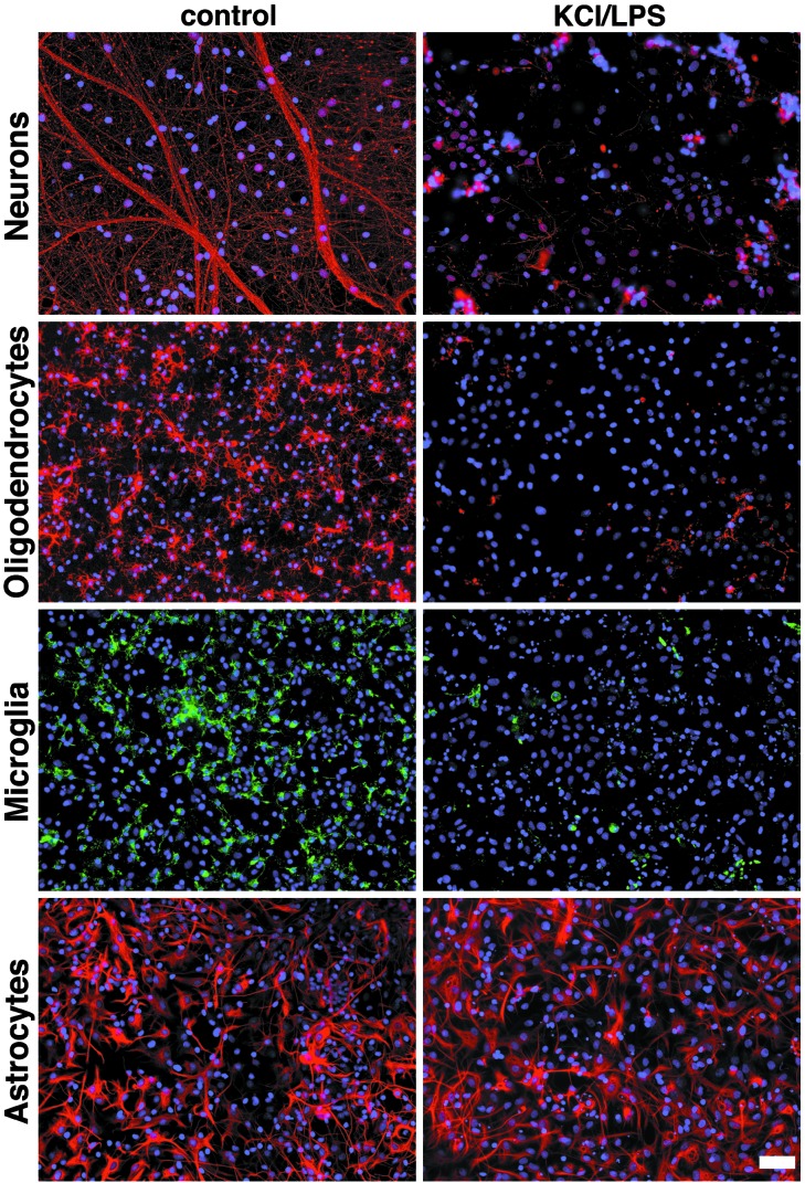
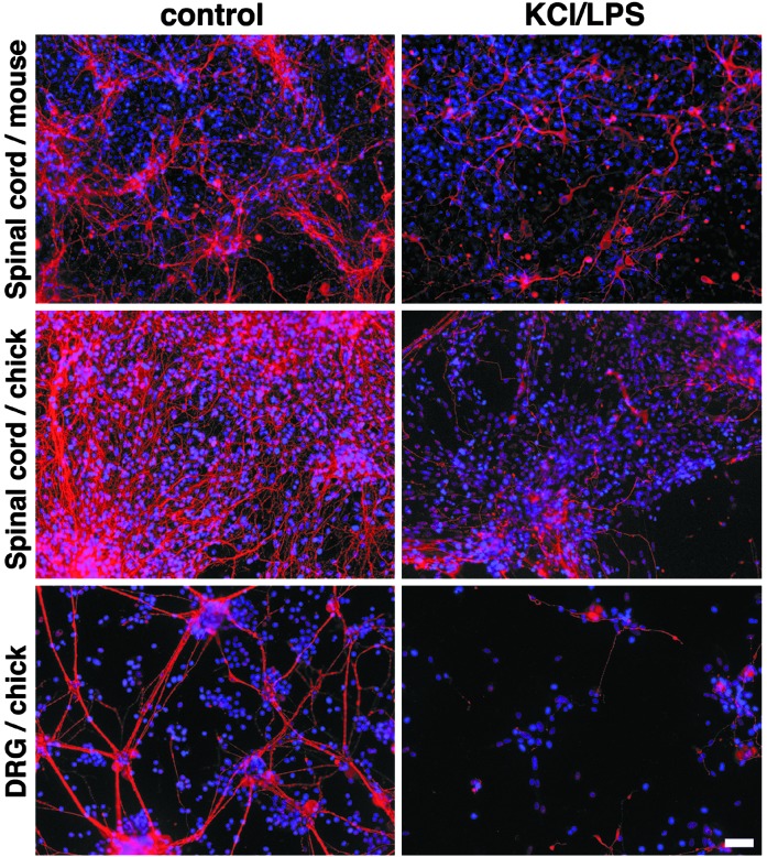
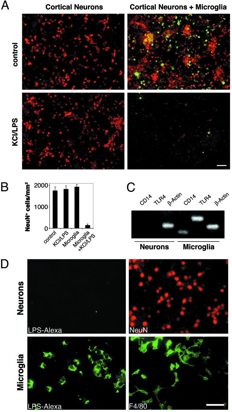
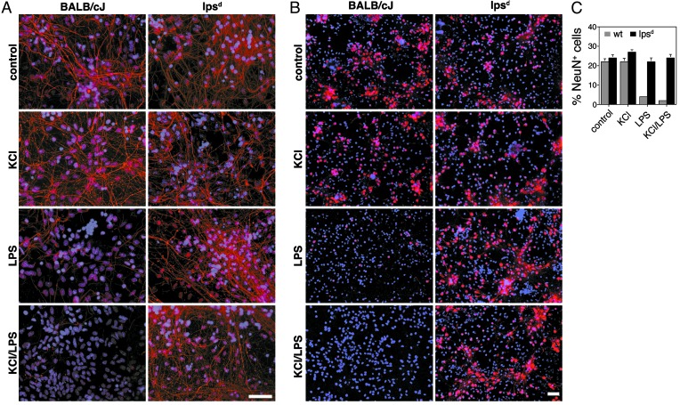
Innate immunity is an evolutionarily ancient system that provides organisms with immediately available defense mechanisms through recognition of pathogen-associated molecular patterns. We show that in the CNS, specific activation of innate immunity through a Toll-like receptor 4 (TLR4)-dependent pathway leads to neurodegeneration. We identify microglia as the major lipopolysaccharide (LPS)-responsive cell in the CNS. TLR4 activation leads to extensive neuronal death in vitro that depends on the presence of microglia. LPS leads to dramatic neuronal loss in cultures prepared from wild-type mice but does not induce neuronal injury in CNS cultures derived from tlr4 mutant mice. In an in vivo model of neurodegeneration, stimulating the innate immune response with LPS converts a subthreshold hypoxic-ischemic insult from no discernable neuronal injury to severe axonal and neuronal loss. In contrast, animals bearing a loss-of-function mutation in the tlr4 gene are resistant to neuronal injury in the same model. The present study demonstrates a mechanistic link among innate immunity, TLRs, and neurodegeneration.


Figures

Fig. 1.
Fig. 1.
LPS induces axonal, oligodendrocyte, and microglial death. Mixed CNS cultures from rat forebrains were incubated with 10 μg/ml LPS in combination with 20 mM KCl or with PBS for 5 days. Staining with antibodies against neurofilament, O4, and glial fibrillary acidic protein marked axons, oligodendrocytes, and astrocytes, respectively. Microglia were stained with isolectin B4. DAPI staining revealed the total number of cells by staining all nuclei. (Scale bar, 50 μm.)
Fig. 2.
Fig. 2.
LPS-induced neurotoxicity does not depend on cell type or species. Mixed CNS cultures were prepared from mouse and chicken spinal cords. Dorsal root ganglion cells (DRG) were generated from chicken, and mixed glia from rat were added. Cultures were treated with 10 μg/ml LPS in combination with 20 mM KCl or PBS for 5 days. Dendrites and axons were stained with microtubule-associated protein 2 and antibody against neurofilament. Nuclei were stained with DAPI. (Scale bar, 50 μm.)
Fig. 3.
Fig. 3.
LPS-induced neurotoxicity is not cell-autonomous but requires microglia. Cortical neurons and microglia were prepared from rat forebrains. (A) Purified neurons and neurons in combination with microglia were incubated with 10 μg/ml LPS/KCl or PBS. After 2 days, cultures were fixed and stained with NeuN and F4/80 to mark neurons and microglia, respectively. (Scale bar, 50 μm.) (B) Quantitation of NeuN+ neurons in purified and microglia-enriched cultures in the presence or absence of LPS/KCl. Results are presented as mean ± SE. P < 0.001. (C) PCR. TLR4 is expressed in microglia but not in neurons. RNA was isolated from purified neuronal and microglial cultures and amplified by RT-PCR with specific primers for TLR4, CD14, or β-actin. (D) Fluorescently labeled LPS binds to microglia but not to neurons. Enriched cultures of microglia and cortical neurons were incubated with Alexa-tagged LPS and analyzed by immunofluorescence. Parallel cultures were stained to identify microglia (F4/80) and neurons (NeuN). (Scale bar, 100 μm.)
Fig. 4.
Fig. 4.
TLR4 is necessary for LPS-induced neuronal injury in vitro. Mixed CNS cultures prepared from BALB/cJ and lpsd mouse forebrains were incubated with 10 μg/ml LPS alone or in combination with 20 mM KCl for 5 days. (A) Cultures were stained with antibody against neurofilament to mark axons and with DAPI. (Scale bar, 50 μm.) (B) Cultures were stained with antibody NeuN (neurons) and DAPI. (Scale bar, 50 μm.) (C) Quantitation of NeuN+ neurons from BALB/cJ and lpsd mice in the presence or absence of LPS. Experiments were repeated three times. Results are presented as mean ± SE. P < 0.001.
Fig. 5.
Fig. 5.
TLR4 is necessary for LPS-induced neuronal injury in vivo. Coronal brain sections of P11 neonatal BALB/cJ and lpsd mice that received a single dose of either LPS or vehicle i.p. before being exposed to HI treatment on P7 were stained with antibodies against neurofilament (A) or NeuN (B). (Scale bar, 50 μm.)

Similar articles

Cited by

References

    1. Kreutzberg, G. W. (1996) Trends Neurosci. 19 312-318. - PubMed
    1. Dickson, D. W., Lee, S. C., Mattiace, L. A., Yen, S. H. & Brosnan, C. (1993) Glia 7 75-83. - PubMed
    1. McGeer, P. L., Itagaki, S., Boyes, B. E. & McGeer, E. G. (1988) Neurology 38 1285-1291. - PubMed
    1. Trapp, B. D., Bo, L., Mork, S. & Chang, A. (1999) J. Neuroimmunol. 98 49-56. - PubMed
    1. Giulian, D., Wendt, E., Vaca, K. & Noonan, C. A. (1993) Proc. Natl. Acad. Sci. USA 90 2769-2773. - PMC - PubMed

Publication types

MeSH terms

Substances

LinkOut - more resources