Review
. 2003 Jul;12(7):1313-22.
doi: 10.1110/ps.0243403.

Crystal structures of fusion proteins with large-affinity tags

Affiliations
Review

Crystal structures of fusion proteins with large-affinity tags

Douglas R Smyth et al. Protein Sci. 2003 Jul.

Abstract

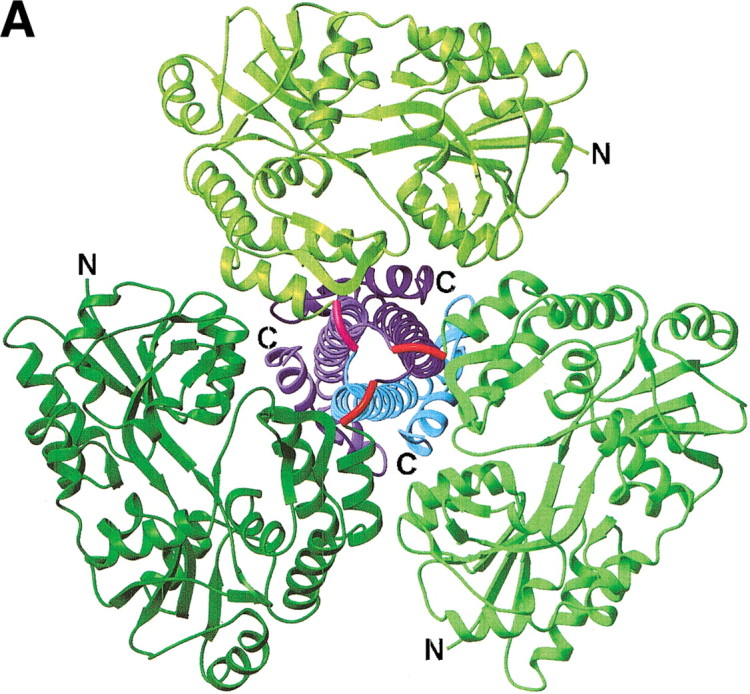
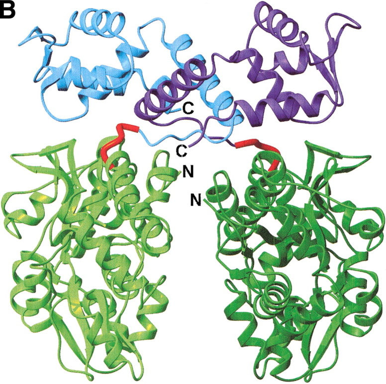
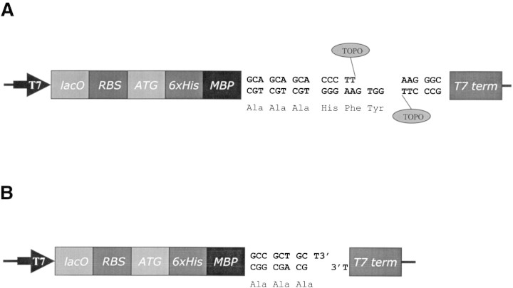
The fusion of a protein of interest to a large-affinity tag, such as the maltose-binding protein (MBP), thioredoxin (TRX), or glutathione-S-transferase (GST), can be advantageous in terms of increased expression, enhanced solubility, protection from proteolysis, improved folding, and protein purification via affinity chromatography. Unfortunately, crystal growth is hindered by the conformational heterogeneity induced by the fusion tag, requiring that the tag is removed by a potentially problematic cleavage step. The first three crystal structures of fusion proteins with large-affinity tags have been reported recently. All three structures used a novel strategy to rigidly fuse the protein of interest to MBP via a short three- to five-amino acid spacer. This strategy has the potential to aid structure determination of proteins that present particular experimental challenges and are not conducive to more conventional crystallization strategies (e.g., membrane proteins). Structural genomics initiatives may also benefit from this approach as a way to crystallize problematic proteins of significant interest.


Figures

Figure 1.
Figure 1.
Structures of MBP fusion proteins. (A) MBP/gp21 trimer. The MBP moieties, the gp21 moieties, and the linkers are shown in different shades of green, blue, and red, respectively. (B) MBP/SarR dimer, shown as in (A). SarR moieties are shown in different shades of blue. (C) MBP/MATa1, shown as in (A). MATa1 moiety is shown in blue. The N- and C-termini are indicated. The figure was prepared with the programs MOLMOL (Koradi et al. 1996) and POV-Ray (http://www.povray.org/).
Figure 1.
Figure 1.
Structures of MBP fusion proteins. (A) MBP/gp21 trimer. The MBP moieties, the gp21 moieties, and the linkers are shown in different shades of green, blue, and red, respectively. (B) MBP/SarR dimer, shown as in (A). SarR moieties are shown in different shades of blue. (C) MBP/MATa1, shown as in (A). MATa1 moiety is shown in blue. The N- and C-termini are indicated. The figure was prepared with the programs MOLMOL (Koradi et al. 1996) and POV-Ray (http://www.povray.org/).
Figure 2.
Figure 2.
(A) Three architectures of membrane protein crystals. (B) A method for growing 2D membrane protein crystals using MBP fusion proteins.
Figure 2.
Figure 2.
(A) Three architectures of membrane protein crystals. (B) A method for growing 2D membrane protein crystals using MBP fusion proteins.
Figure 3.
Figure 3.
(A) Schematic diagram of the proposed TOPO expression vector, incorporating the MBP affinity tag, three alanines, and a TOPO directional cloning site. T7, T7 promoter; lacO, lac operon; RBS, ribosome binding site; ATG, translation initiation codon; 6xHis, His-tag; MBP, maltose binding protein; T7 term, T7 termination region; TOPO, topoisomerase I. (B) Schematic diagram of the proposed nondirectional expression vector, incorporating the MBP affinity tag and three alanines, labeled as in (A). 3’ T overhangs are indicated.

Similar articles

Cited by

References

    1. Abramson, J. and Iwata, S. 1999. Crystallization of membrane proteins. In Protein crystallization: Techniques, strategies, and tips: A laboratory manual (ed. T.M. Bergfors), pp. 199–210. International University Line, La Jolla, CA.
    1. Baneyx, F. 1999. Recombinant protein expression in Escherichia coli. Curr. Opin. Biotechnol. 10 411–421. - PubMed
    1. Beckwith, J. 2000. The all purpose gene fusion. Methods Enzymol. 326 3–7. - PubMed
    1. Boos, W. and Shuman, H. 1998. Maltose/maltodextrin system of Escherichia coli: Transport, metabolism, and regulation. Microbiol. Mol. Biol. Rev. 62 204. - PMC - PubMed
    1. Bornhorst, J.A. and Falke, J.J. 2000. Purification of proteins using polyhistidine affinity tags. Methods Enzymol. 326 245–254. - PMC - PubMed

Publication types

MeSH terms