Review
. 2001 Sep;4(5):561-74.

G protein-coupled receptor drug discovery: implications from the crystal structure of rhodopsin

Affiliations
Review

G protein-coupled receptor drug discovery: implications from the crystal structure of rhodopsin

J Ballesteros et al. Curr Opin Drug Discov Devel. 2001 Sep.

Abstract

G protein-coupled receptors (GPCRs) are a functionally diverse group of membrane proteins that play a critical role in signal transduction. Because of the lack of a high-resolution structure, the heptahelical transmembrane bundle within the N-terminal extracellular and C-terminal intracellular region of these receptors has initially been modeled based on the high-resolution structure of bacterial retinal-binding protein, bacteriorhodopsin. However, the low-resolution structure of rhodopsin, a prototypical GPCR, revealed that there is a minor relationship between GPCRs and bacteriorhodopsins. The high-resolution crystal structure of the rhodopsin ground state and further refinements of the model provide the first structural information about the entire organization of the polypeptide chain and post-translational moieties. These studies provide a structural template for Family 1 GPCRs that has the potential to significantly improve structure-based approaches to GPCR drug discovery.


Figures

Figure 1
Figure 1
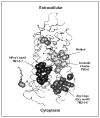
Helical net representation of the transmembrane domain of rhodopsin, showing the most conserved residues across Family 1 GPCRs. Solvent accessible residues within the TM domain, proposed to face lipids, are shaded in gray. Highly conserved residues within Family 1 GPCRs are marked with bold font and a thick circle. Residues within the top half of the TM domain not facing the lipid milieu form the binding site crevice accessible to extracellular ligands. A family-based numbering scheme is utilized, preceded by the TM number and followed by its position within the TM segment relative to the most conserved and recognizable residue within each TM, which is denoted 1.50, 2.50, etc.
Figure 2
Figure 2
Conserved residues in rhodopsin 3D structure: Functional microdomains. The highly conserved residues identified in Figure 1 are shown in a view parallel to the membrane of the rhodopsin structure. These set of residues cluster in 3D, defining three distinct functional microdomains: retinal is in direct contact with the Aromatic Cluster on the TM5-TM6 interface, which is adjacent to the NPxxY motif that characterizes TM7 and includes a polar pocket composed by N1.50-D2.50-N7.49 in TM1-2-7-8. Residues surrounding the conserved DRY motif in TM3 near the cytoplasmic boundary form the Arg-Cage microdomain. The conserved disulfide bond on the extracellular side and the palmitoylation site at the end of TM8 on the cytoplasmic side are shown.
Figure 3
Figure 3
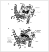
Divergent structures for the extracellular segment of GPCRs: Crystal structures from different GPCRs. Comparison of the crystal structure of the extracellular domain of rhodopsin (A) [12●●] with the ectodomain of Methusalah (B) (a G protein from the secretin receptor family) [80], and the N-terminal domain of a metabotropic glutamate receptor (C) [81●].
Figure 4
Figure 4
The residues in the TM domain of rhodopsin in direct contact with retinal comprise the most important ligand binding residues identified for other GPCRs. (A) Extracellular view showing the residues comprising the binding site for retinal within the TM domain of rhodopsin. (B) Analogous view of a D4-selective compound (CPPMA) docked onto a model of the dopamine D4 receptor derived from the rhodopsin structure based on gain of function mutagenesis. The key interaction residues for dopamine (D3.32, S5.42, S5.43, S5.46, W6.48, F6.52) and the residues that confer selective recognition of CPPMA relative to the D2 receptor subtype (F2.61, L3.28, M3.29, V7.35) are shown. Note the overlap between the binding site crevices of both receptors. All identified contact residues in the D4/D2 receptors occupy positions that are in direct contact with retinal in rhodopsin.
Figure 5
Figure 5
Epinephrine and β2 -adrenergic receptor. (A) Molecular model based on the rhodopsin structure of epinephrine (EPI) binding to the β2-adrenergic receptor. Docking of the ligand was based on specific ligand-receptor interactions identified in the last 12 years by correlating minor modifications on the ligand with site-directed receptor mutagenesis. The protonated amine of EPI interacts with D3.32 in TM3, the catelcholamine hydroxyls hydrogen bond serine residues S5.42, S5.43 and S5.46 in TM5, the aromatic moiety of EPI interacts with W6.48 and F6.52 in TM6, and the hydroxyl interacts with N6.52 in TM6. (B) Alternative orientations of the phenyl moiety of EPI resulting from hydrogen bonding of catelcholamine hydroxyls to S5.43 and S5.46 in TM5 as reported originally, or to S5.42, S5.43, and S5.46 as reported more recently. (C) Alternative orientations of the phenyl moiety of EPI resulting from hydrogen bonding of catelcholamine hydroxyls to S5.42, S5.43 and S5.46 in TM5 in two different rotamer orientations of the S5.46 side chain: the gauge- rotamer conformation of S5.46 shown also in Panel B is compared to the gauge+ conformation of S5.46. Note the significant spatial reorientation of positions suitable for chemical modification as required to optimize a lead compound, which would interact with different residues within the receptor's binding site.
Figure 6
Figure 6
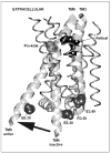
Similar conformational changes upon receptor activation in rhodopsin and other GPCRs: TM3-TM6. View parallel to the membrane plane of the rhodopsin structure highlighting TM3 and TM6 (large ribbons). In the crystallized inactive state, TM6 is close to TM3 at the cytoplasmic boundary, held by an 'ionic lock' between R3.50 and E3.49-E6.30, the Arg-Cage motif in Figure 2. Experimental data on both rhodopsin and multiple receptors (see text) have shown that upon receptor activation, these acidic residues become protonated releasing the 'ionic lock' and TM6 moves away from TM3 (black arrow). The conserved Pro-kink motif in TM6 may function as a flexible hinge responsible for the observed TM6 movement, shifting from a kinked helix (inactive) to a straighter helix (active) (black arrow). The Pro-kink motif belongs to the aromatic cluster (see Figure 2) at the level of the ligand binding site, shown here by retinal. Light-driven isomerization of retinal would trigger conformational changes in the aromatic cluster microdomain that relieve the Pro-kink motif, which will propagate the signal across the transmembrane. The result is a more flexible TM6 that, on average, is further away from TM3, opening the cytoplasmic domain for G protein coupling. The shared binding site and activation mechanism between rhodopsin and these other GPCRs suggests that the resulting active state structure should also be similar.
Scheme 1
Scheme 1
Ligand-activated and rhodopsin GPCRs.

References

    1. Sautel M, Milligan G. Molecular manipulation of G-protein-coupled receptors: A new avenue into drug discovery. Curr Med Chem. 2000;7:889–896. - PubMed
    1. Muller G. Towards 3D structures of G protein-coupled receptors: A multidisciplinary approach. Curr Med Chem. 2000;7:861–888. ● This review summarizes the recent results on GPCR structural studies. - PubMed
    1. Bargmann CI. Neurobiology of the Caenorhabditis elegans genome. Science. 1998;282:2028–2033. - PubMed
    1. Larhammar D, Blomqvist AG, Wahlestedt C. The receptor revolution - multiplicity of G-protein-coupled receptors. Drug Des Discov. 1993;9:179–188. - PubMed
    1. Howard AD, McAllister G, Feighner SD, Liu Q, Nargund RP, Van der Ploeg LH, Patchett AA. Orphan G-protein-coupled receptors and natural ligand discovery. Trends Pharmacol Sci. 2001;22:132–140. - PubMed

Publication types

LinkOut - more resources